Difference between revisions of "ProjectWeek200706:LesionClassificationInLupus"
From NAMIC Wiki
Hjbockholt (talk | contribs) |
Hjbockholt (talk | contribs) |
||
| Line 30: | Line 30: | ||
Our approach is to utilize existing automatic lesion classification techniques, both within NA-MIC kit and external to it. | Our approach is to utilize existing automatic lesion classification techniques, both within NA-MIC kit and external to it. | ||
| − | Our plan for the project week is to learn and use EMSegment (S. Wells), BRAINS2 (V. Magnotta), and Constructing Image Graphs for Lesion Segmention (M. Prastawa) | + | Our plan for the project week is to learn and use EMSegment (S. Wells), BRAINS2 (V. Magnotta), and Constructing Image Graphs for Lesion Segmention (M. Prastawa). We will learn and begin to compare and contrast these approaches with a bronze standard (manual rater tracings) of lesions. |
</div> | </div> | ||
Revision as of 11:56, 25 June 2007
Home < ProjectWeek200706:LesionClassificationInLupus Return to Project Week Main Page |
Key Investigators
- MIND/UNM:
- Jeremy Bockholt
- Mark Scully
- Core 1
- Bruce Fischl
- External Collaborator
- Vincent Magnotta
Objective
Our goal is to automatically, or with little or no manual human rater input, accurately tissue classify our example lupus data-set into gray, white, csf, and lesion classes.
Approach, Plan
Our approach is to utilize existing automatic lesion classification techniques, both within NA-MIC kit and external to it.
Our plan for the project week is to learn and use EMSegment (S. Wells), BRAINS2 (V. Magnotta), and Constructing Image Graphs for Lesion Segmention (M. Prastawa). We will learn and begin to compare and contrast these approaches with a bronze standard (manual rater tracings) of lesions.
Progress
This section will get filled out at the end of the Project week.
References
Additional Information
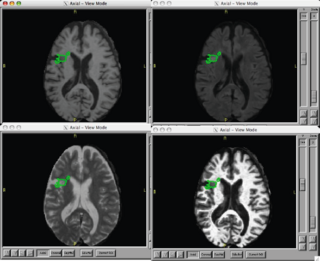
Using an exemplar case that has already been processed using non-NAMIC kit tools:
- Use existing NA-MIC kit to coregister T1, T2, Flair
- Use the EM Segment in slicer 2-3.X to classify grey, white, csf, and white matter lesion
- Summarize the volume and location of lesions
