Difference between revisions of "2008 Winter Project Week:MRISC"
| Line 1: | Line 1: | ||
| + | {| | ||
| + | |[[Image:NAMIC-SLC.jpg|thumb|320px|Return to [[2008_Winter_Project_Week]] ]] | ||
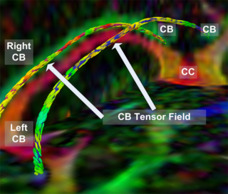
| + | |valign="top"|[[Image:Case24-coronal-tensors-edit.png |thumb|320px|The Cingulum Bundle Anchor Tract]] | ||
| + | |} | ||
| − | + | __NOTOC__ | |
| + | ===Key Investigators=== | ||
| + | * MIT CSAIL: Mert R Sabuncu | ||
| + | * BWH: Kilian Pohl | ||
| − | This study will explore an approach that utilizes an atlas-based joint | + | <div style="margin: 20px;"> |
| − | registration segmentation framework to automatically classify | + | |
| − | magnetic resonance images into healthy and disease subjects. We | + | <div style="width: 27%; float: left; padding-right: 3%;"> |
| − | extend an Expectation Maximization framework (Pohl et al. 2006) that | + | |
| − | unifies atlas-based segmentation and registration. The geometric | + | <h1>Objective</h1> |
| − | transformation (i.e., registration with the atlas) is defined using | + | This study will explore an approach that utilizes an atlas-based joint registration segmentation framework to automatically classify |
| − | structure-specific affine parameters that yield a global, one-to-one | + | magnetic resonance images into healthy and disease subjects. We extend an Expectation Maximization framework (Pohl et al. 2006) that unifies atlas-based segmentation and registration. The geometric transformation (i.e., registration with the atlas) is defined using structure-specific affine parameters that yield a global, one-to-one non-linear deformation. The modes of variation of the transformation parameters are generated using principal component analysis. This provides a robust way to learn the global deformation space that aligns a new brain with the atlas. With this parametrization, the algorithm then performs a deformation-based classification of the new brain using a Support Vector Machine. We demonstrate the algorithm with a data set that consists of 16 first episode schizophrenics and 17 healthy subjects. The data set included manual labels for the (left and right) Superior Temporal Gyrus (STG), Hippocampus (HIPP), Amygdala (AMY) and Parahippocampal Gyrus (PHG). |
| − | non-linear deformation. The modes of variation of the transformation | + | |
| − | parameters are generated using principal component analysis. This | + | </div> |
| − | provides a robust way to learn the global deformation space that | + | |
| − | aligns a new brain with the atlas. With this parametrization, the | + | <div style="width: 27%; float: left; padding-right: 3%;"> |
| − | algorithm then performs a deformation-based classification of the | + | |
| − | new brain using a Support Vector Machine. We demonstrate the | + | <h1>Approach, Plan </h1> |
| − | algorithm with a data set that consists of 16 first episode | + | Our approach is builds on the technology described in the reference below. |
| − | schizophrenics and 17 healthy subjects. The data set | + | </div> |
| − | included manual labels for the (left and right) Superior Temporal | + | |
| − | Gyrus (STG), Hippocampus (HIPP), Amygdala (AMY) and Parahippocampal | + | <div style="width: 40%; float: left;"> |
| − | Gyrus (PHG). | + | |
| + | <h1>Progress</h1> | ||
| + | |||
| + | ====Jan 2008 Project Half Week==== | ||
| + | |||
| + | |||
| + | </div> | ||
| + | |||
| + | <br style="clear: both;" /> | ||
| + | |||
| + | </div> | ||
| + | |||
| + | |||
| + | ===Reference=== | ||
| + | |||
| + | K. M. Pohl, J. Fisher, W.E.L. Grimson, R. Kikinis, and W.M. Wells. A bayesian model for joint segmentation and registration. NeuroImage, 31(1), pp. 228-239, 2006 | ||
Revision as of 19:38, 14 December 2007
Home < 2008 Winter Project Week:MRISC Return to 2008_Winter_Project_Week |
Key Investigators
- MIT CSAIL: Mert R Sabuncu
- BWH: Kilian Pohl
Objective
This study will explore an approach that utilizes an atlas-based joint registration segmentation framework to automatically classify magnetic resonance images into healthy and disease subjects. We extend an Expectation Maximization framework (Pohl et al. 2006) that unifies atlas-based segmentation and registration. The geometric transformation (i.e., registration with the atlas) is defined using structure-specific affine parameters that yield a global, one-to-one non-linear deformation. The modes of variation of the transformation parameters are generated using principal component analysis. This provides a robust way to learn the global deformation space that aligns a new brain with the atlas. With this parametrization, the algorithm then performs a deformation-based classification of the new brain using a Support Vector Machine. We demonstrate the algorithm with a data set that consists of 16 first episode schizophrenics and 17 healthy subjects. The data set included manual labels for the (left and right) Superior Temporal Gyrus (STG), Hippocampus (HIPP), Amygdala (AMY) and Parahippocampal Gyrus (PHG).
Approach, Plan
Our approach is builds on the technology described in the reference below.
Progress
Jan 2008 Project Half Week
Reference
K. M. Pohl, J. Fisher, W.E.L. Grimson, R. Kikinis, and W.M. Wells. A bayesian model for joint segmentation and registration. NeuroImage, 31(1), pp. 228-239, 2006
