Difference between revisions of "EM Segmentation For Orthopaedic Applications"
From NAMIC Wiki
| Line 15: | Line 15: | ||
'''Results:''' | '''Results:''' | ||
| − | Finger Phalanx Segment | + | {| class="wikitable" border="1" cellpadding="2" |
| − | Index (14 subjects) Proximal 0.87 | + | |+Relative Overlap with Manual Rater |
| − | + | |- | |
| − | + | ! Finger !! Phalanx Segment !! Average Relative Overlap | |
| − | + | |- | |
| − | + | ! Index (14 subjects) | |
| − | + | | Proximal || 0.87 | |
| − | Middle (2 subjects) Proximal 0.79 | + | |- |
| − | + | ! | |
| − | + | | Medial || 0.80 | |
| − | Ring (2 subjects) Proximal 0.76 | + | |- |
| − | + | ! | |
| − | + | | Distal || 0.70 | |
| − | Pinky (2 subjects) Proximal 0.70 | + | |- |
| − | + | ! Middle (2 subjects) | |
| − | + | | Proximal || 0.79 | |
| + | |- | ||
| + | ! | ||
| + | | Medial || 0.77 | ||
| + | |- | ||
| + | ! | ||
| + | | Distal || 0.71 | ||
| + | |- | ||
| + | ! Ring (2 subjects) | ||
| + | | Proximal || 0.76 | ||
| + | |- | ||
| + | ! | ||
| + | | Medial || 0.82 | ||
| + | |- | ||
| + | ! | ||
| + | | Distal || 0.80 | ||
| + | |- | ||
| + | ! Pinky (2 subjects) | ||
| + | | Proximal || 0.70 | ||
| + | |- | ||
| + | ! | ||
| + | | Medial || 0.73 | ||
| + | |- | ||
| + | ! | ||
| + | | Distal || 0.72 | ||
| + | |} | ||
| + | |||
'''To Do:''' | '''To Do:''' | ||
Revision as of 00:07, 7 January 2008
Home < EM Segmentation For Orthopaedic ApplicationsObjective:
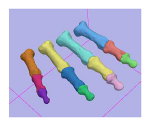
- To utilize the Slicer3 Expectation Maximization Algorithm for segmentation of the phalanx bones of the hand.
Progress:
- We have utilized the Slicer2.7 EM Segmentation Module for segmentation of the phalanx bones
- Registration was performed outside of Slicer using ITK registration algorithms that are available in the IaFeMesh software. This includes Thin plate spline, B-Spline and rigid registration algorithms.
- Probability map information was created from a single subject used as the atlas image and filtered using a Gaussian filter.
- Initial evaluation has been performed
- Reliability assessed on fourteen specimens that also had the index finger segmented, and two specimens that had the index, middle, ring and little fingers manually segmented
- Validation assessed using the laser scanning of the index finger (proximal, middle, and distal) phalanx bones in five specimens
- Work is underway to develop a Slicer2.7 tutorial for this segmentation (File:Draft EM Segment Tutorial 9 12.pdf)
Results:
| Finger | Phalanx Segment | Average Relative Overlap |
|---|---|---|
| Index (14 subjects) | Proximal | 0.87 |
| Medial | 0.80 | |
| Distal | 0.70 | |
| Middle (2 subjects) | Proximal | 0.79 |
| Medial | 0.77 | |
| Distal | 0.71 | |
| Ring (2 subjects) | Proximal | 0.76 |
| Medial | 0.82 | |
| Distal | 0.80 | |
| Pinky (2 subjects) | Proximal | 0.70 |
| Medial | 0.73 | |
| Distal | 0.72 |
To Do:
- Evaluate the Slicer3 EM Segmentation Module
- Example datasets sent to Brad Davis for evaluation of the Slicer3 EM Segmentation for non-neuro applications
- Discussion of the results will take place at 2008 AHM
- Update the the tutorial to support the Slicer3 Workflow
- Summer of 2008 - Tutorial will be developed by Austin Ramme
- Determine if registration tools in Slicer are adequate for this work.
Key Investigators:
- Iowa: Austin Ramme, Nicole Grosland, and Vincent Magnotta
Links:
Figures:

