Difference between revisions of "Integration of Neural Network Algorithms"
From NAMIC Wiki
| (6 intermediate revisions by the same user not shown) | |||
| Line 19: | Line 19: | ||
* Iowa: Nicole DeVries, Nicole Grosland, Vincent Magnotta | * Iowa: Nicole DeVries, Nicole Grosland, Vincent Magnotta | ||
| + | |||
| + | |||
| + | '''Results''' | ||
| + | {| class="wikitable" border="1" cellpadding="2" | ||
| + | |+ANN Relative Overlap with Manual Rater | ||
| + | |- | ||
| + | ! Subject !! Proximal Phalanx !! Middle Phalanx !! Distal Phalanx | ||
| + | |- | ||
| + | | 1 || 0.91 || 0.79 || 0.79 | ||
| + | |- | ||
| + | | 2 || 0.91 || 0.88 || 0.84 | ||
| + | |- | ||
| + | | 3 || 0.85 || 0.83 || 0.78 | ||
| + | |- | ||
| + | | 4 || 0.86 || 0.81 || 0.68 | ||
| + | |- | ||
| + | | 5 || 0.84 || 0.78 || 0.72 | ||
| + | |- | ||
| + | ! Average ||0.87 || 0.82 || 0.76 | ||
| + | |} | ||
| + | |||
| + | <br> | ||
| + | <br> | ||
| + | |||
| + | {| class="wikitable" border="1" cellpadding="2" | ||
| + | |+Average Distance Between ANN Derived Surface and Physical Laser Scanned Surface | ||
| + | |- | ||
| + | ! Subject !! Proximal Phalanx (mm) !! Middle Phalanx (mm) !! Distal Phalanx (mm) | ||
| + | |- | ||
| + | | 1 || 0.23||0.12||0.17 | ||
| + | |- | ||
| + | | 2 || 0.18||0.16||0.16 | ||
| + | |- | ||
| + | | 3 || 0.35||0.27||0.97 | ||
| + | |- | ||
| + | | 4 || 0.26||0.17||0.20 | ||
| + | |- | ||
| + | ! Bone Average ||0.26||0.18||0.38 | ||
| + | |} | ||
| + | |||
'''Links:''' | '''Links:''' | ||
*[http://www.springerlink.com/content/b15376vwx00v5088/ Automated Bony Region Identification Using Artificial Neural Networks: Reliability and Validation Measurements. Esther E. Gassman, Stephanie M. Powell, Nicole A. Kallemeyn, Kiran H. Shivanna, Vincent A. Magnotta, Brian D. Adams, Nicole M. Grosland. Skeletal Radiology] | *[http://www.springerlink.com/content/b15376vwx00v5088/ Automated Bony Region Identification Using Artificial Neural Networks: Reliability and Validation Measurements. Esther E. Gassman, Stephanie M. Powell, Nicole A. Kallemeyn, Kiran H. Shivanna, Vincent A. Magnotta, Brian D. Adams, Nicole M. Grosland. Skeletal Radiology] | ||
| + | *[http://www.ccad.uiowa.edu/mimx Musculoskeletal Imaging, Modeling and Experimentation (MIMX)] | ||
| + | |||
| + | '''Figures:''' | ||
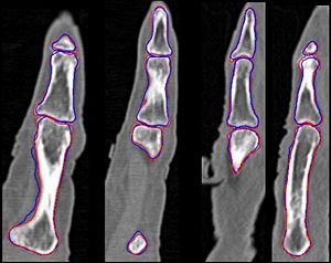
| + | [[Image:ANN-Segmentation-Phalanx.jpg |left|thumb|300px|Comparison of the manual and ANN segmentations of the phalanx bones. Manual segmentations are shown in red and automated in blue.]] | ||
Latest revision as of 00:40, 7 January 2008
Home < Integration of Neural Network AlgorithmsObjective:
- Integrate artificial neural network segmentation into the NA-MIC kit for segmentation of upper extremity regions of interest.
Progress:
- Base algorithm has been developed using the annie neural network library
- Segmentation algorithm has been applied to segmentation of the phalanx bones on the index finger
- Paper has been published on this initial work
To Do:
- Initial evaluation of this technique has been performed for phalanx bones.
- Need to rework ITK neural network libraries to allow the size of the network to be dynamic configured at run time instead of compile time
- There are several issues that neeed to be resolved regarding the Neural network I/O in ITK
- Consider the inclusion of the old ANN code that existed in the BRAINS software
Key Investigators:
- Iowa: Nicole DeVries, Nicole Grosland, Vincent Magnotta
Results
| Subject | Proximal Phalanx | Middle Phalanx | Distal Phalanx |
|---|---|---|---|
| 1 | 0.91 | 0.79 | 0.79 |
| 2 | 0.91 | 0.88 | 0.84 |
| 3 | 0.85 | 0.83 | 0.78 |
| 4 | 0.86 | 0.81 | 0.68 |
| 5 | 0.84 | 0.78 | 0.72 |
| Average | 0.87 | 0.82 | 0.76 |
| Subject | Proximal Phalanx (mm) | Middle Phalanx (mm) | Distal Phalanx (mm) |
|---|---|---|---|
| 1 | 0.23 | 0.12 | 0.17 |
| 2 | 0.18 | 0.16 | 0.16 |
| 3 | 0.35 | 0.27 | 0.97 |
| 4 | 0.26 | 0.17 | 0.20 |
| Bone Average | 0.26 | 0.18 | 0.38 |
Links:
- Automated Bony Region Identification Using Artificial Neural Networks: Reliability and Validation Measurements. Esther E. Gassman, Stephanie M. Powell, Nicole A. Kallemeyn, Kiran H. Shivanna, Vincent A. Magnotta, Brian D. Adams, Nicole M. Grosland. Skeletal Radiology
- Musculoskeletal Imaging, Modeling and Experimentation (MIMX)
Figures:
