Difference between revisions of "Projects/Diffusion/2008 Project Week DiffusionMRI QBall"
| (2 intermediate revisions by the same user not shown) | |||
| Line 2: | Line 2: | ||
|[[Image:ProjectWeek-2008.png|thumb|320px|Return to [[2008_Programming/Project_Week_MIT|Project Week Main Page]] ]] | |[[Image:ProjectWeek-2008.png|thumb|320px|Return to [[2008_Programming/Project_Week_MIT|Project Week Main Page]] ]] | ||
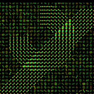
|[[Image:SynQBall.png|thumb|320px|Synthetical Q-Ball Image.]] | |[[Image:SynQBall.png|thumb|320px|Synthetical Q-Ball Image.]] | ||
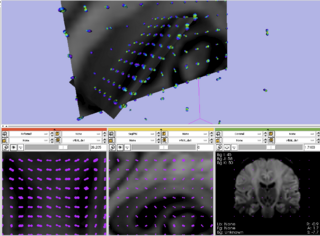
| − | [[Image:SlicerQBall.png|thumb|320px|Slicer showing Q-Ball Image.]] | + | |[[Image:SlicerQBall.png|thumb|320px|Slicer showing a Q-Ball Image.]] |
|} | |} | ||
| Line 31: | Line 31: | ||
<div style="width: 40%; float: left;"> | <div style="width: 40%; float: left;"> | ||
| + | |||
| + | <h1>Progress</h1> | ||
| + | |||
| + | Glyph visualization for volume images in different slices was successfully improved in order to show QBall and other diffusion MRI images. The QBall image visualization was successfully improved. However, the current architecture depends on the three slice model (Red, Yellow, Green) and is too specific for the Diffusion MRI case. Efforts are currently taking place in order to solve these two drawbacks. | ||
</div> | </div> | ||
Latest revision as of 10:32, 27 June 2008
Home < Projects < Diffusion < 2008 Project Week DiffusionMRI QBall Return to Project Week Main Page |
Key Investigators
- Odyssée, INRIA, France: Demian Wassermann, Rachid Deriche
- LMI, Brigham & Womens Hospital, Harvard Medical School: CF Westin
Objective
We are using the currently in development Sliced Multiple Layer Glyph visualization for Volume data to Q-Ball visualization into Slicer3.
Approach, Plan
Our approach is to add a new MRML Volume type with the needed MRML display modules and widgets. Probably, some of the the MRML sctructure for Diffusion Image representation and visualization will need minor modifications along with the Logic and GUI packages of Slicer3 in order to allow easy integration of new glyphed visualization schemes.
Our plan for the project is to end with a working prototype,...
Progress
Glyph visualization for volume images in different slices was successfully improved in order to show QBall and other diffusion MRI images. The QBall image visualization was successfully improved. However, the current architecture depends on the three slice model (Red, Yellow, Green) and is too specific for the Diffusion MRI case. Efforts are currently taking place in order to solve these two drawbacks.
References
- Basser, P. & Pierpaoli, C. Microstructural and physiological features of tissues elucidated by quantitative-diffusion-tensor MRI Journal of Magnetic Resonance B, 1996, 111, 209-219
- Westin, C.; Maier, S. E.; Mamata, H.; Nabavi, A.; Jolesz, F. A. & Kikinis, R. Processing and visualization for diffusion tensor MRI. Med Image Anal, 2002, 6, 93-108
- Tuch, D. S. Q-ball imaging, Magnetic Resonance in Medicine, 2004, 52, 1358-1372
- Lenglet, C.; Rousson, M.; Deriche, R. & Faugueras, O. Statistics on the Manifold of Multivariate Normal Distributions: Theory and Application to Diffusion Tensor MRI Processing J Math Imaging Vis, 2006, 25, 423-444
- Descoteaux, M.; Angelino, E.; Fitzgibbons, S. & Deriche, R. Apparent Diffusion Coefficients from High Angular Resolution Diffusion Images: Estimation and Applications, Magnetic Resonance in Medicine, 2006, 56, 395-410

