Difference between revisions of "2009 Winter Project Week:LocalCorticalThicknessPipeline"
From NAMIC Wiki
(New page: {| |thumb|320px|Return to [[2009_Winter_Project_Week|Project Week Main Page ]] |} __NOTOC__ ===Key Investigators=== * UNC: Clement Vachet, Martin Styner, Heathe...) |
|||
| (2 intermediate revisions by the same user not shown) | |||
| Line 1: | Line 1: | ||
{| | {| | ||
|[[Image:NAMIC-SLC.jpg|thumb|320px|Return to [[2009_Winter_Project_Week|Project Week Main Page]] ]] | |[[Image:NAMIC-SLC.jpg|thumb|320px|Return to [[2009_Winter_Project_Week|Project Week Main Page]] ]] | ||
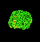
| + | |[[Image:CorticalThickness_WhiteMatter.jpg|thumb|170px| Regional cortical thickness analysis on white matter surface]] | ||
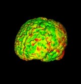
| + | |[[Image:CorticalThickness_GreyMatter.jpg|thumb|170px|Regional cortical thickness analysis on grey matter surface]] | ||
|} | |} | ||
| Line 8: | Line 10: | ||
===Key Investigators=== | ===Key Investigators=== | ||
| − | * UNC: Clement Vachet, Martin Styner, Heather Cody Hazlett, Marc Niethammer | + | * UNC: Clement Vachet, Martin Styner, Heather Cody Hazlett, Marc Niethammer, Ipek Oguz |
* GE: Jim Miller | * GE: Jim Miller | ||
| Line 28: | Line 30: | ||
<h1>Approach, Plan</h1> | <h1>Approach, Plan</h1> | ||
| − | Our plan for the project week is to | + | Our plan for the project week is to improve the pipeline and focus on the following steps: |
| + | *Mesh inflation with mesh correction | ||
| + | *Inflated mesh correspondence using particle systems | ||
| + | |||
| + | We will perform the analysis on a small dataset. | ||
| + | |||
</div> | </div> | ||
| Line 35: | Line 42: | ||
<h1>Progress</h1> | <h1>Progress</h1> | ||
| − | + | The local cortical thickness pipeline has been slightly modified to include WMMap fixing | |
| + | *The mesh inflation step has been updated to detect vertices that have a high curvature (extremities) | ||
| + | *A new module has been developped to correct the WMMap image | ||
| + | *Pipeline loops until the fixing is no longer necessary | ||
| + | |||
| + | |||
| + | Meetings (with Martin, Dan Marcus, Wendy Plesniak) and concerning future integration of the tool within Slicer3: use of XNAT for group analysis. | ||
</div> | </div> | ||
Latest revision as of 17:02, 9 January 2009
Home < 2009 Winter Project Week:LocalCorticalThicknessPipeline Return to Project Week Main Page |
Key Investigators
- UNC: Clement Vachet, Martin Styner, Heather Cody Hazlett, Marc Niethammer, Ipek Oguz
- GE: Jim Miller
Objective
We are developing an end-to-end application within Slicer3 allowing individual and group analysis of local cortical thickness.
Such a workflow applied to the young brain (2-4 years old) is our goal in order to start a longitudinal study of early brain development in autism (UNC DBP).
See our Roadmap for more details.
Approach, Plan
Our plan for the project week is to improve the pipeline and focus on the following steps:
- Mesh inflation with mesh correction
- Inflated mesh correspondence using particle systems
We will perform the analysis on a small dataset.
Progress
The local cortical thickness pipeline has been slightly modified to include WMMap fixing
- The mesh inflation step has been updated to detect vertices that have a high curvature (extremities)
- A new module has been developped to correct the WMMap image
- Pipeline loops until the fixing is no longer necessary
Meetings (with Martin, Dan Marcus, Wendy Plesniak) and concerning future integration of the tool within Slicer3: use of XNAT for group analysis.

