Difference between revisions of "Slicer3.2:Training"
| Line 34: | Line 34: | ||
| style="background:#C3D1C3; color:black" align="Center"| [[Image:SceneRestore.png|200px|LoadingandVisualization]] | | style="background:#C3D1C3; color:black" align="Center"| [[Image:SceneRestore.png|200px|LoadingandVisualization]] | ||
|- | |- | ||
| − | | style="background:#9BF2C5; color:black" align="Center"| '''1.2''' | + | | style="background:#9BF2C5; color:black" align="Center"| <span id="1.2"></span> '''1.2''' |
| style="background:#D1FFF9; color:black"| [[Media:Manual Segmentation with 3DSlicer-mj.ppt| Manual segmentation with 3D Slicer]] | | style="background:#D1FFF9; color:black"| [[Media:Manual Segmentation with 3DSlicer-mj.ppt| Manual segmentation with 3D Slicer]] | ||
| style="background:#D1FFF9; color:black"| | | style="background:#D1FFF9; color:black"| | ||
| style="background:#C3D1C3; color:black" align="Center"| [[Image:OrbitSegmentationt.png|200px]] | | style="background:#C3D1C3; color:black" align="Center"| [[Image:OrbitSegmentationt.png|200px]] | ||
|- | |- | ||
| − | | style="background:#9BF2C5; color:black" align="Center"| '''1.3''' | + | | style="background:#9BF2C5; color:black" align="Center"| <span id="1.3"></span> '''1.3''' |
| style="background:#D1FFF9; color:black"| [[Media:AutomaticSegmentation_SoniaPujol_Munich2008.ppt|EM Segmentation Course]]<br> | | style="background:#D1FFF9; color:black"| [[Media:AutomaticSegmentation_SoniaPujol_Munich2008.ppt|EM Segmentation Course]]<br> | ||
Background Materials:[[EMSegmenter| EMSegmenter History]] | Background Materials:[[EMSegmenter| EMSegmenter History]] | ||
| Line 48: | Line 48: | ||
| style="background:#C3D1C3; color:black" align="Center"| [[Image:EMSegmentation2008.png|200px|EM Segmenter]] | | style="background:#C3D1C3; color:black" align="Center"| [[Image:EMSegmentation2008.png|200px|EM Segmenter]] | ||
|- | |- | ||
| − | | style="background:#9BF2C5; color:black" align="Center"| '''1.4''' | + | | style="background:#9BF2C5; color:black" align="Center"| <span id="1.4"></span> '''1.4''' |
| style="background:#D1FFF9; color:black"|[[Media:Na-MIC-Slicer-Registration.ppt|Affine and Deformable Registration in Slicer 3]] | | style="background:#D1FFF9; color:black"|[[Media:Na-MIC-Slicer-Registration.ppt|Affine and Deformable Registration in Slicer 3]] | ||
| style="background:#D1FFF9; color:black"| [[Media:SlicerSampleRegistration.tgz | SlicerSampleRegistration.tgz]]<br> | | style="background:#D1FFF9; color:black"| [[Media:SlicerSampleRegistration.tgz | SlicerSampleRegistration.tgz]]<br> | ||
| Line 54: | Line 54: | ||
| style="background:#C3D1C3; color:black" align="Center"| [[Image:Na-MIC-Slicer-Registration.png|200px|SlicerRegistration]] | | style="background:#C3D1C3; color:black" align="Center"| [[Image:Na-MIC-Slicer-Registration.png|200px|SlicerRegistration]] | ||
|- | |- | ||
| − | | style="background:#9BF2C5; color:black" align="Center"| '''1.5''' | + | | style="background:#9BF2C5; color:black" align="Center"| <span id="1.5"></span> '''1.5''' |
| style="background:#D1FFF9; color:black"| [[Media:slicer3diffusionTutorial7.pdf| Processing of Diffusion Weighted Imaging and Diffusion Tensor Imaging data in Slicer3]]<br> | | style="background:#D1FFF9; color:black"| [[Media:slicer3diffusionTutorial7.pdf| Processing of Diffusion Weighted Imaging and Diffusion Tensor Imaging data in Slicer3]]<br> | ||
Background Materials:[http://slicer.org/slicerWiki/index.php/Slicer3:DTMRI DT-MRI module]. | Background Materials:[http://slicer.org/slicerWiki/index.php/Slicer3:DTMRI DT-MRI module]. | ||
| Line 60: | Line 60: | ||
| style="background:#C3D1C3; color:black" align="Center"| [[Image:Line-glyph-tracts.jpg|200px|Glyphs and tracts]] | | style="background:#C3D1C3; color:black" align="Center"| [[Image:Line-glyph-tracts.jpg|200px|Glyphs and tracts]] | ||
|- | |- | ||
| − | | style="background:#9BF2C5; color:black" align="Center"| '''1.6''' | + | | style="background:#9BF2C5; color:black" align="Center"| <span id="1.6"></span> '''1.6''' |
| style="background:#D1FFF9; color:black"| [[Media:Slicer3Training ChangeTracker.ppt| Detecting subtle change in pathology]] <br> | | style="background:#D1FFF9; color:black"| [[Media:Slicer3Training ChangeTracker.ppt| Detecting subtle change in pathology]] <br> | ||
This tutorial consists of two MR scans of a patient with meningioma. | This tutorial consists of two MR scans of a patient with meningioma. | ||
| Line 66: | Line 66: | ||
| style="background:#C3D1C3; color:black" align="Center"| [[Image:TumorGrowth-Tutorial-Image.png|200px|Meningioma Case]] | | style="background:#C3D1C3; color:black" align="Center"| [[Image:TumorGrowth-Tutorial-Image.png|200px|Meningioma Case]] | ||
|- | |- | ||
| − | | style="background:#9BF2C5; color:black" align="Center"| '''1.7''' | + | | style="background:#9BF2C5; color:black" align="Center"| <span id="1.7"></span> '''1.7''' |
| style="background:#D1FFF9; color:black"| [http://www.nitrc.org/frs/download.php/516/Slicer3Training_WhiteMatterLesions_1.1.ppt.pdf Detecting white matter lesions in lupus] <br> | | style="background:#D1FFF9; color:black"| [http://www.nitrc.org/frs/download.php/516/Slicer3Training_WhiteMatterLesions_1.1.ppt.pdf Detecting white matter lesions in lupus] <br> | ||
This tutorial consists of five MR scans of patients with lupus. | This tutorial consists of five MR scans of patients with lupus. | ||
| Line 73: | Line 73: | ||
| style="background:#C3D1C3; color:black" align="Center"| [[Image:Lupus2.png|200px|Lesion Segmentation]] | | style="background:#C3D1C3; color:black" align="Center"| [[Image:Lupus2.png|200px|Lesion Segmentation]] | ||
|- | |- | ||
| − | | style="background:#9BF2C5; color:black" align="Center"| '''1.8''' | + | | style="background:#9BF2C5; color:black" align="Center"| <span id="1.8"></span> '''1.8''' |
| style="background:#D1FFF9; color:black"| [[Media:FreeSurferCourse_SoniaPujol.ppt|FreeSurfer Course]] <br> | | style="background:#D1FFF9; color:black"| [[Media:FreeSurferCourse_SoniaPujol.ppt|FreeSurfer Course]] <br> | ||
This tutorial teaches how to visualize FreeSurfer segmentations and parcellation maps of the brain. | This tutorial teaches how to visualize FreeSurfer segmentations and parcellation maps of the brain. | ||
| Line 79: | Line 79: | ||
| style="background:#C3D1C3; color:black" align="Center"| [[Image:FreeSurferParcellation.PNG|200px|FreeSurfer segmentation]] | | style="background:#C3D1C3; color:black" align="Center"| [[Image:FreeSurferParcellation.PNG|200px|FreeSurfer segmentation]] | ||
|- | |- | ||
| − | | style="background:#8EDEB5; color:black" align="Center"| '''2.1''' | + | | style="background:#8EDEB5; color:black" align="Center"| <span id="2.1"></span> '''2.1''' |
| style="background:#D1FFF9; color:black"| [[media:ProgrammingIntoSlicer3_SoniaPujol.ppt |Programming into Slicer3]] | | style="background:#D1FFF9; color:black"| [[media:ProgrammingIntoSlicer3_SoniaPujol.ppt |Programming into Slicer3]] | ||
This tutorial is intended for engineers and scientists who want to integrate stand-alone programs outside of the Slicer3 source tree. | This tutorial is intended for engineers and scientists who want to integrate stand-alone programs outside of the Slicer3 source tree. | ||
| Line 86: | Line 86: | ||
| style="background:#C3D1C3; color:black" align="Center"| [[Image:ProgrammingCourse_Logo.PNG|200px|Programming]] | | style="background:#C3D1C3; color:black" align="Center"| [[Image:ProgrammingCourse_Logo.PNG|200px|Programming]] | ||
|- | |- | ||
| − | | style="background:#8EDEB5; color:black" align="Center"| '''2.2''' | + | | style="background:#8EDEB5; color:black" align="Center"| <span id="2.2"></span> '''2.2''' |
| style="background:#D1FFF9; color:black"| [[Media:ImageGuidedTherapyPlanning.pdf|Image Guided Therapy Planning Tutorial]]<br> | | style="background:#D1FFF9; color:black"| [[Media:ImageGuidedTherapyPlanning.pdf|Image Guided Therapy Planning Tutorial]]<br> | ||
This tutorial takes the trainee through a complete workup for neurosurgical patient specific mapping.See pages 58 to 80 of this tutorial on using the '''"Simple region growing"''' segmentation module. | This tutorial takes the trainee through a complete workup for neurosurgical patient specific mapping.See pages 58 to 80 of this tutorial on using the '''"Simple region growing"''' segmentation module. | ||
| Line 93: | Line 93: | ||
| style="background:#C3D1C3; color:black" align="Center"| [[Image:NeurosurgicalPlanningOverview.png|200px|Neurosurgical Planning Overview]] | | style="background:#C3D1C3; color:black" align="Center"| [[Image:NeurosurgicalPlanningOverview.png|200px|Neurosurgical Planning Overview]] | ||
|- | |- | ||
| − | | style="background:#72b291; color:black" align="Center"| '''3.1''' | + | | style="background:#72b291; color:black" align="Center"| <span id="3.1"></span> '''3.1''' |
| style="background:#D1FFF9; color:black"| [http://wiki.na-mic.org/Wiki/index.php/IGT:ToolKit Slicer3 as a research tool for image guided therapy research (IGT)] | | style="background:#D1FFF9; color:black"| [http://wiki.na-mic.org/Wiki/index.php/IGT:ToolKit Slicer3 as a research tool for image guided therapy research (IGT)] | ||
This tutorial is intended for engineers and scientists who want to use Slicer 3 for IGT research. | This tutorial is intended for engineers and scientists who want to use Slicer 3 for IGT research. | ||
| Line 99: | Line 99: | ||
| style="background:#C3D1C3; color:black" align="Center"| [[Image:Slicer IGTL NITRobot.jpg|200px|Slicer with robots]] | | style="background:#C3D1C3; color:black" align="Center"| [[Image:Slicer IGTL NITRobot.jpg|200px|Slicer with robots]] | ||
|- | |- | ||
| − | | style="background:#72b291; color:black" align="Center"| '''3.2''' | + | | style="background:#72b291; color:black" align="Center"| <span id="3.2"></span> '''3.2''' |
| style="background:#D1FFF9; color:black"| [[Media:Brain Atlas Tutorial08.ppt| SPL-PNL Brain Atlas Tutorial]] | | style="background:#D1FFF9; color:black"| [[Media:Brain Atlas Tutorial08.ppt| SPL-PNL Brain Atlas Tutorial]] | ||
This three-dimensional brain atlas dataset, derived from a volumetric, whole-head MRI scan, contains the original MRI-scan, a complete set of label maps, three-dimensional reconstructions (200+ structures) and a tutorial. <br> | This three-dimensional brain atlas dataset, derived from a volumetric, whole-head MRI scan, contains the original MRI-scan, a complete set of label maps, three-dimensional reconstructions (200+ structures) and a tutorial. <br> | ||
| Line 106: | Line 106: | ||
| style="background:#C3D1C3; color:black" align="Center"| [[Image:Atlas.png|200px|Brain Atlas]] | | style="background:#C3D1C3; color:black" align="Center"| [[Image:Atlas.png|200px|Brain Atlas]] | ||
|- | |- | ||
| − | | style="background:#72b291; color:black" align="Center"| '''3.3''' | + | | style="background:#72b291; color:black" align="Center"| <span id="3.3"></span> '''3.3''' |
| style="background:#D1FFF9; color:black"| [[Media:Abdominal_Atlas_Tutorial08.ppt| SPL Abdominal Atlas Tutorial]] | | style="background:#D1FFF9; color:black"| [[Media:Abdominal_Atlas_Tutorial08.ppt| SPL Abdominal Atlas Tutorial]] | ||
This three-dimensional abdominal atlas was derived from a computed tomography (CT) scan, using semi-automated image segmentation and three-dimensional reconstruction techniques. The dataset contains the original CT scan, detailed label maps, three-dimensional reconstructions and a tutorial. | This three-dimensional abdominal atlas was derived from a computed tomography (CT) scan, using semi-automated image segmentation and three-dimensional reconstruction techniques. The dataset contains the original CT scan, detailed label maps, three-dimensional reconstructions and a tutorial. | ||
| Line 113: | Line 113: | ||
| style="background:#C3D1C3; color:black" align="Center"| [[Image:Abdominal-Atlas-3Dsnapshot.png|200px|Abdominal Atlas]] | | style="background:#C3D1C3; color:black" align="Center"| [[Image:Abdominal-Atlas-3Dsnapshot.png|200px|Abdominal Atlas]] | ||
|- | |- | ||
| − | | style="background:#72b291; color:black" align="Center"| '''3.4''' | + | | style="background:#72b291; color:black" align="Center"| <span id="3.4"></span> '''3.4''' |
| style="background:#D1FFF9; color:black"| [http://slicer.spl.harvard.edu/slicerWiki/index.php/Slicer3:XND Using XNAT Desktop and Slicer3 for remote data handling ] | | style="background:#D1FFF9; color:black"| [http://slicer.spl.harvard.edu/slicerWiki/index.php/Slicer3:XND Using XNAT Desktop and Slicer3 for remote data handling ] | ||
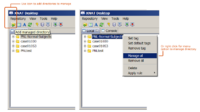
This tutorial is intended for researchers who wish to upload local datasets to a remote XNAT repository like [http://central.xnat.org '''XNAT Central'''] and retrieve catalog files that can be opened inside Slicer. | This tutorial is intended for researchers who wish to upload local datasets to a remote XNAT repository like [http://central.xnat.org '''XNAT Central'''] and retrieve catalog files that can be opened inside Slicer. | ||
| Line 119: | Line 119: | ||
| style="background:#C3D1C3; color:black" align="Center"| [[Image:XNDmanage2.png|200px|XNAT Desktop Manager]] | | style="background:#C3D1C3; color:black" align="Center"| [[Image:XNDmanage2.png|200px|XNAT Desktop Manager]] | ||
|- | |- | ||
| − | | style="background:#72b291; color:black" align="Center"| '''3.5''' | + | | style="background:#72b291; color:black" align="Center"| <span id="3.5"></span> '''3.5''' |
| style="background:#D1FFF9; color:black"| '''Harnessing Grid Computational Resources and integration with Slicer through the Grid Wizard Enterprise system''' | | style="background:#D1FFF9; color:black"| '''Harnessing Grid Computational Resources and integration with Slicer through the Grid Wizard Enterprise system''' | ||
These tutorials are intended for researchers who wish to utilize distributed computational resources. | These tutorials are intended for researchers who wish to utilize distributed computational resources. | ||
Revision as of 20:49, 6 April 2009
Home < Slicer3.2:TrainingWelcome to the 3D Slicer3.2 User Training 101
The information on this page applies to 3D Slicer version 3. If you are looking for materials about Slicer version 2, please visit the Slicer2 101 page.
|
Slicer Logo | |
Software Installation
The Slicer download page contains links for downloading the different versions of Slicer 3.
Training Compendium
| Category | Tutorial | Sample Data | Image |
| 1.1 | Data Loading and 3D Visualization in Slicer3 | SlicerSampleVisualization.tar.gz SlicerSampleVisualization.zip |
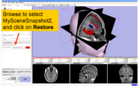
|
| 1.2 | Manual segmentation with 3D Slicer | 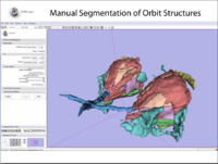
| |
| 1.3 | EM Segmentation Course Background Materials: EMSegmenter History |
AutomaticSegmentation.tar.gz
|
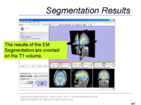
|
| 1.4 | Affine and Deformable Registration in Slicer 3 | SlicerSampleRegistration.tgz |
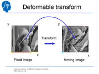
|
| 1.5 | Processing of Diffusion Weighted Imaging and Diffusion Tensor Imaging data in Slicer3 Background Materials:DT-MRI module. |
Slicer3DiffusionTutorialData.zip |
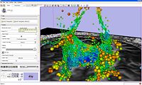
|
| 1.6 | Detecting subtle change in pathology This tutorial consists of two MR scans of a patient with meningioma. |
ChangeTracker-Tutorial-Data.zip | 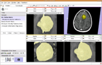
|
| 1.7 | Detecting white matter lesions in lupus This tutorial consists of five MR scans of patients with lupus. |
Lupus-Tutorial-Data.tgz |

|
| 1.8 | FreeSurfer Course This tutorial teaches how to visualize FreeSurfer segmentations and parcellation maps of the brain. |
FreeSurfer Tutorial Data FreeSurferTutorialData.zip |
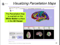
|
| 2.1 | Programming into Slicer3
This tutorial is intended for engineers and scientists who want to integrate stand-alone programs outside of the Slicer3 source tree. |
HelloWorld_Plugin.zip | 
|
| 2.2 | Image Guided Therapy Planning Tutorial This tutorial takes the trainee through a complete workup for neurosurgical patient specific mapping.See pages 58 to 80 of this tutorial on using the "Simple region growing" segmentation module.
|
NeurosurgicalPlanningTutorialData.zip | 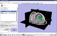
|
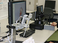
| 3.1 | Slicer3 as a research tool for image guided therapy research (IGT)
This tutorial is intended for engineers and scientists who want to use Slicer 3 for IGT research. |
| |
| 3.2 | SPL-PNL Brain Atlas Tutorial
This three-dimensional brain atlas dataset, derived from a volumetric, whole-head MRI scan, contains the original MRI-scan, a complete set of label maps, three-dimensional reconstructions (200+ structures) and a tutorial. |
SPL-PNL Brain Atlas | 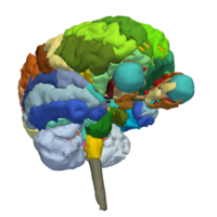
|
| 3.3 | SPL Abdominal Atlas Tutorial
This three-dimensional abdominal atlas was derived from a computed tomography (CT) scan, using semi-automated image segmentation and three-dimensional reconstruction techniques. The dataset contains the original CT scan, detailed label maps, three-dimensional reconstructions and a tutorial.
|
SPL Abdominal Atlas | 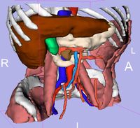
|
| 3.4 | Using XNAT Desktop and Slicer3 for remote data handling
This tutorial is intended for researchers who wish to upload local datasets to a remote XNAT repository like XNAT Central and retrieve catalog files that can be opened inside Slicer. |
| |
| 3.5 | Harnessing Grid Computational Resources and integration with Slicer through the Grid Wizard Enterprise system
These tutorials are intended for researchers who wish to utilize distributed computational resources. |
Grid Wizard Enterprise | 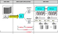 Featured Study Case (YouTube) Featured Study Case (YouTube)
|
- Category 1 = Basic functionality
- Category 2 = Advanced functionality
- Category 3 = Specialized application packages
Additional Materials
For a variety of data sets for downloading, check the following link.
Back to Training:Main
