Difference between revisions of "DBP2:Queens:Roadmap"
Sidd queens (talk | contribs) |
Sidd queens (talk | contribs) |
||
| Line 16: | Line 16: | ||
The primary goal for the roadmap is to develop an interventional module for Slicer3 for MRI-guided prostate biopsies. This module and the accompanying tutorial will serve as a template for interventional applications with Slicer3. The module will provide the necessary functionality for calibrating the robot to the MR scanner, planning biopsies, computing the necessary robot trajectory to perform each biopsy, and verification via post-biopsy images. We will obtain a biopsy plan from multi-parametric endorectal image volumes, executable with an existing prostate biopsy device. The system will be will be implemented under Slicer3 as an interactive application. | The primary goal for the roadmap is to develop an interventional module for Slicer3 for MRI-guided prostate biopsies. This module and the accompanying tutorial will serve as a template for interventional applications with Slicer3. The module will provide the necessary functionality for calibrating the robot to the MR scanner, planning biopsies, computing the necessary robot trajectory to perform each biopsy, and verification via post-biopsy images. We will obtain a biopsy plan from multi-parametric endorectal image volumes, executable with an existing prostate biopsy device. The system will be will be implemented under Slicer3 as an interactive application. | ||
| + | |||
<center> | <center> | ||
{| | {| | ||
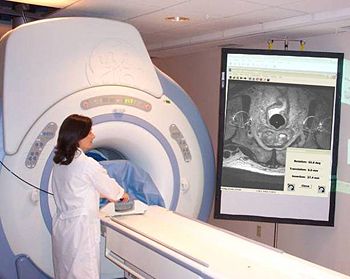
| − | |valign="top"|[[Image: | + | |valign="top"|[[Image:Menard.jpg|thumb|350px|Prostate intervention (biopsy) in closed MR scanner.]] |
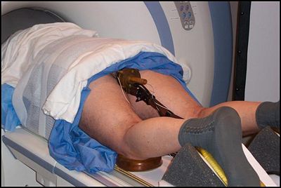
| + | |[[Image:Patient.jpg|Close-up of the transrectal procedure|thumb|400px]] | ||
|} | |} | ||
</center> | </center> | ||
| + | |||
| + | <center> | ||
| + | {| | ||
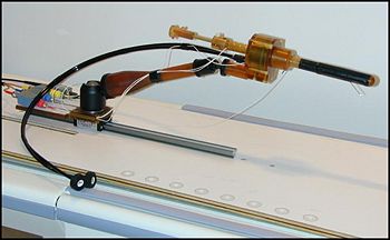
| + | |valign="top"|[[Image:Robot.jpg|thumb|350px|Transrectal prostate intervention robot assembled.]] | ||
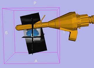
| + | |[[Image:TRProstateBiopsyRobot.jpg|thumb|320px|The transrectal prostate robot.]] | ||
| + | |} | ||
| + | </center> | ||
| + | |||
== '''Current status''' == | == '''Current status''' == | ||
Revision as of 17:15, 29 April 2009
Home < DBP2:Queens:RoadmapBack to NA-MIC Internal Collaborations, JHU DBP 2
Objective
We would like to create an end-to-end application within the NA-MIC Kit to enable an existing transrectal prostate biopsy device to perform multi-parametric MRI guided prostate biopsy in closed-bore high-field MRI magnets.
This page describes the technology roadmap for robotic prostate biopsy in the NA-MIC Kit. The basic components necessary for this application are:
- Tissue segmentation: Should be multi-modality, correcting for intensity inhomogeneity and work for both supine and prone patients, all imaged with an endorectal coil (ERC).
- Registration: co-registration of MRI datasets taken at different times, in different body positions, and under different imaging parameters
- Prostate Measurement: Measure volume of all segmented structures
- Biopsy Device Parameters: Geometry, kinematics, and calibration/registration of the robot system must be available in some form. This capability is not currently part of the NA-MIC kit. The application will be modular, to enable use of different devices.
- Tutorial: Documentation will be written for a tutorial and sample data sets will be provided to perform simulated biopsies.
Roadmap
The primary goal for the roadmap is to develop an interventional module for Slicer3 for MRI-guided prostate biopsies. This module and the accompanying tutorial will serve as a template for interventional applications with Slicer3. The module will provide the necessary functionality for calibrating the robot to the MR scanner, planning biopsies, computing the necessary robot trajectory to perform each biopsy, and verification via post-biopsy images. We will obtain a biopsy plan from multi-parametric endorectal image volumes, executable with an existing prostate biopsy device. The system will be will be implemented under Slicer3 as an interactive application.
Current status
- Segmentation: Semi-automatic segmentation has been implemented by Fichtinger et al. Statistical atlas based segmentation has been prototyped by Tannenbaum et al. Pose-independent segmentation (workable in both supine/prone) is being implemented.
- Registration: contour based registration has been prototyped by Fichtinger et al., needs to be re-implemented with native NA-MIC tools.
- Prostate Measurement: Prototyped by Fichtinger et al., needs to be re-implemented with native NA-MIC tools.
- Device Modeling: Prototyped by Fichtinger et al, needs to be re-implemented with native NA-MIC tools.
- Biopsy Planning: Clinically functional, currently being implemented with native NA-MIC tools
- Tutorial: Not yet started. It will be derived from the actual clinically-functional system, with demo data.
Schedule
Data Collection: Done. Initial data available, hand segmented for ground truth.
Segmentation: We plan to use shape-based segmentation methods for the MRI prostate data. Several parts of the procedure have already been implemented with NA-MIC tools such as the conformal flattening procedure. Spherical wavelets for shape analysis are already available in ITK. Despeckling techniques will be used to enhance ultrasound imagery as a pre-processing step for segmentation of the prostate data.
System Implementation: Apart from the one research element (segmentation), the rest of the project is a massive software engineering effort, and will follow these major milestones and schedule:
Application Workflow Development:
10/15/2007 Define the workflow for the application (David, Csaba, Gabor) --- DONE ---
10/22/2007 Create GUI templates for the workflow steps only till Calibration step (David) --- DONE ---
11/20/2008 Wizard GUI created for Targeting step, and Verification step (Siddharth) --- DONE ---
Software architecture compliance of module:
11/20/2008 Model View Controller pattern reflected in corresponding MRML, GUI, Logic (Siddharth) ---DONE---
Device Modeling:
12/15/2007 Conversion of engineering data into VTK-viewable objects (David) -- DONE --
Data Display:
12/15/2007 Provide display logic for targets and prostate outlines (David) --- DONE ---
Measurement Tools:
07/01/2008 Semi-automatic identification of fiducials via thresholding & centroids still not integrated inside SLICER (Csaba) --- DONE ---
11/26/2008 Integration of semi-automatic identification of fiducials in the SLICER module (Siddharth) ---DONE---
07/01/2008 Logic for robot registration with fiducials still not integrated inside SLICER (Csaba) --- DONE ---
11/26/2008 Integration of Logic for robot registration with fiducials in the SLICER module --- IN PROCESS ---
08/01/2008 Prostate measurement tools (David) --- REMOVED (use automatic segmentation instead) ---
Biopsy Planning/Targeting:
07/01/2008 Calculations for robot trajectory based on target position still not integrated inside SLICER (Csaba) --- DONE ---
11/24/2008 Integration of calculations for robot trajectory based on target position (Siddharth) --- IN PROCESS ---
11/24/2008 Implement planning tools (display, logic) in Slicer3 (Siddharth, David) --- IN PROCESS ---
09/19/2008 Verification against pre-existing software and data (David, Siddharth)
Robot positioning:
09/19/2008 GUI targeting readouts for optical encoders (David, Siddharth)---- TO BE DONE----
11/10/2008 Integrate optical encoders with our Slicer module (Siddharth) -----DONE------
Integration of Prostate segmentation (by Yi, Tennanbaum) in SLICER module:
01/05/2009 - 01/09-2009 Planned integration of segmentation developed at Georgia Tech by Yi, Tennanbaum during the NAMIC project week in Utah --- PLANNED ACTIVITY --- The current status and schedule is here: prostate segmentation AHM2009
Verification and testing of integrated system:
Software & documentation
- The TRProstateBiopsy module is in the "Queens" directory of the NAMICSandBox - access online
Tutorial (end-to-end):
- Trans-rectal prosate biopsy module: TransRectalProstateBiopsyTutorial
- Trans-rectal prostate biopsy tutorial dataset: TRPBTutorialDataset.zip
Publications
- Krieger A, Susil RC, Menard C, Coleman JA, Fichtinger G, Atalar E, Whitcomb LL, Design of A Novel MRI Compatible Manipulator for Image Guided Prostate Intervention, IEEE Trans. Biomed. Eng. 2005; 52(2):306-313
- Susil RC, Ménard C, Krieger A, Coleman JA, Camphausen K, Choyke P, Ullman K, Smith S, Fichtinger G, Whitcomb LL, Coleman NC, Atalar E, Transrectal Prostate Biopsy and Fiducial Marker Placement in a Standard 1.5T MRI Scanner, J Urol. 2006 Jan;175(1):113-20
- GS Fischer, A Krieger, I Iordachita, LL Whitcomb, and G Fichtinger, "MRI Compatibility of Robot Actuation Techniques -- A Comparative Study", Eleventh International Conference on Medical Image Computing and Computer-Assisted Intervention (MICCAI), Proceedings in Lecture Notes in Computer Science Vol. 5242, pp 509-517, Springer, 2008
- Siddharth Vikal, Steven Haker, Clare Tempany, Gabor Fichtinger, Prostate contouring in MRI guided biopsy, SPIE Medical Imaging 2009
- S. Gill, P Abolmaesumi, S Vikal, P Mousavi, G. Fichtinger, Intraoperative Prostate Tracking with Slice-to-Volume Registration in MRI, 20th International Conference of the Society for Medical Innovation and Technology (SMIT), Vienna, Austria, August 28-31, 2008, Electronic proceedings, ISBN 3-902087-25-0, pp. 154-158
- Mewes PW, Tokuda J, DiMaio SP, Fischer GS, Csoma C, Gobbi DG, Tempany CM, Fichtinger G, Hata N, An Integrated MRI and Robot Control Software for an MRI compatible Robot in Prostate Intervention, International Conference on Robotics and Automation - ICRA 2008, Pasadena, CA, pp 2950-2962, May 2008
Team and Institutes
- PI: Gabor Fichtinger, Queen’s University (gabor at cs.queensu.ca)
- Co-I: Purang Abolmaesumi, Queen’s University (purang at cs.queensu.ca)
- Software Engineer Lead: Siddharth Vikal, Queen’s University (vikal at cs.queensu.ca
- Software Engineer: David Gobbi, Queen’s University (dgobbi at cs.queensu.ca)
- JHU Software Engineer Support: Csaba Csoma, Johns Hopkins University, csoma at jhu.edu
- NA-MIC Engineering Contact: Katie Hayes, MSc, Brigham and Women's Hospital, hayes at bwh.harvard.edu
- NA-MIC Algorithms Contact: Allen Tannenbaum, PhD, GeorgiaTech, tannenba at ece.gatech.edu
- Host Institutes: Queen's University & Johns Hopkins University


