Difference between revisions of "User:Haehn"
| Line 6: | Line 6: | ||
! Info || | ! Info || | ||
|- style="vertical-align:top" | |- style="vertical-align:top" | ||
| − | |'''Daniel Hähn'''<br>Student of Medical Informatics <br>University of Heidelberg, Germany<br>E-Mail: [mailto:haehn@bwh.harvard.edu haehn@bwh.harvard.edu] or [mailto:haehn@urz.uni-heidelberg.de haehn@urz.uni-heidelberg.de]<br>Expected Graduation: | + | |'''Daniel Hähn'''<br>Student of Medical Informatics <br>University of Heidelberg, Germany<br>E-Mail: [mailto:haehn@bwh.harvard.edu haehn@bwh.harvard.edu] or [mailto:haehn@urz.uni-heidelberg.de haehn@urz.uni-heidelberg.de]<br>Expected Graduation: Spring 2010 || <center>[[Image:haehn.jpg]]</center> |
|} | |} | ||
| Line 13: | Line 13: | ||
'''Project goal:''' Integration of VMTK in 3D Slicer<br> | '''Project goal:''' Integration of VMTK in 3D Slicer<br> | ||
'''Kick-Off:''' 10/15/2008 | '''Kick-Off:''' 10/15/2008 | ||
| − | ''' | + | '''Milestone 1:''' 03/31/2009 |
The extraction of vessels in two and threedimensional images is part of many clinical analysis tasks. | The extraction of vessels in two and threedimensional images is part of many clinical analysis tasks. | ||
Revision as of 10:30, 25 November 2009
Daniel Haehn's Page
| Info | |
|---|---|
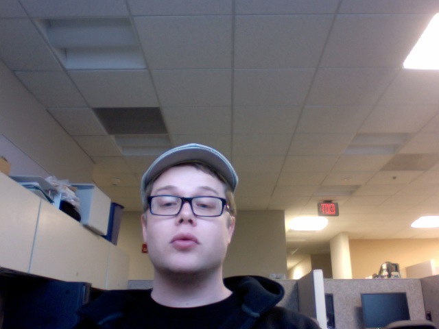
| Daniel Hähn Student of Medical Informatics University of Heidelberg, Germany E-Mail: haehn@bwh.harvard.edu or haehn@urz.uni-heidelberg.de Expected Graduation: Spring 2010 |
 |
VMTK in 3D Slicer
Project goal: Integration of VMTK in 3D Slicer
Kick-Off: 10/15/2008
Milestone 1: 03/31/2009
The extraction of vessels in two and threedimensional images is part of many clinical analysis tasks.
Surgical and radiology procedures often involve the visualization and quantification of vessels in order
to perform surgical planning or diagnostics. There is no single segmentation method that can extract
vessels from every medical image modality, but different approaches and robust algorithms exist.
Various published key algorithms are available within an opensource framework for imagebased
modeling of blood vessels, referred to as the Vascular Modeling Toolkit (VMTK).
The library of VMTK was made available in 3D Slicer, an application providing a wide range of tools
for medical image processing. This was realized using a hidden loadable module approach in order to
provide a flexible way of distributing and including the library. To evaluate and verify the integration, a
software module offering VMTK level set segmentation methods within 3D Slicer was created.
With the successful connection of the two above mentioned software solutions, processing pipelines
between VMTK code and other algorithms can be established. Several techniques for three dimensional
reconstruction, geometric analysis, mesh generation and surface data analysis for imagebased modeling
of blood vessels are now accessible to the 3D Slicer developer. The reference implementation for
accessing VMTK, as well as the created library module are available as opensource software.
The NITRC project page including a SVN repository with the latest code: http://www.nitrc.org/projects/slicervmtklvlst/
More recent news on this project:
http://www.na-mic.org/Wiki/index.php/2009_Winter_Project_Week_Slicer_VMTK
http://www.na-mic.org/Wiki/index.php/Summer2009:The_Vascular_Modeling_Toolkit_in_3D_Slicer
VMTK is the Vascular Modeling Toolkit (http://vmtk.org) and offers interesting techniques for segmentation of vessels or tube-shapes.
This work is done in conjunction with Luca Antiga (Medical Imaging Unit, Bioengineering Department, Mario Negri Institute, Bergamo, Italy).