Difference between revisions of "2010 Winter Project Week Spine Segmentation Module in Slicer3"
Sylvainjaume (talk | contribs) |
Sylvainjaume (talk | contribs) |
||
| Line 3: | Line 3: | ||
Image:spine_segmentation_module_in_slicer_001.png|Interface of the SpineSegmentation module in Slicer 3.5 | Image:spine_segmentation_module_in_slicer_001.png|Interface of the SpineSegmentation module in Slicer 3.5 | ||
Image:spine_segmentation_module_in_slicer_002.png|Results of the SpineSegmentation module in Slicer 3.5 | Image:spine_segmentation_module_in_slicer_002.png|Results of the SpineSegmentation module in Slicer 3.5 | ||
| − | Image:spine_segmentation_module_in_slicer_004.png|3D triangle mesh and tetrahedra mesh of our segmentation results</gallery> | + | Image:spine_segmentation_module_in_slicer_004.png|3D triangle mesh and tetrahedra mesh of our segmentation results |
| + | Image:spine-segmentation-slicer-cylinder.png|Double cylinder model used for the segmentation of the spine | ||
| + | </gallery> | ||
==Key Investigators== | ==Key Investigators== | ||
| Line 44: | Line 46: | ||
{| | {| | ||
| − | |[[Image: | + | |[[Image:spine_segmentation_in_slicer_004.png|thumb|280px|Meshes of our automated segmentation of the spine]] |
| − | | | ||
|[[Image:spine_segmentation_module_in_slicer_000.png|thumb|280px|Results of the SpineSegmentation module in Slicer 3.5]] | |[[Image:spine_segmentation_module_in_slicer_000.png|thumb|280px|Results of the SpineSegmentation module in Slicer 3.5]] | ||
|[[Image:spine_segmentation_module_in_slicer_003.png|thumb|280px|Results of the SpineSegmentation module in Slicer 3.5]] | |[[Image:spine_segmentation_module_in_slicer_003.png|thumb|280px|Results of the SpineSegmentation module in Slicer 3.5]] | ||
Revision as of 07:16, 13 January 2010
Home < 2010 Winter Project Week Spine Segmentation Module in Slicer3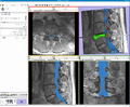
Interface of the SpineSegmentation module in Slicer 3.5
Results of the SpineSegmentation module in Slicer 3.5
- Spine segmentation module in slicer 004.png
3D triangle mesh and tetrahedra mesh of our segmentation results

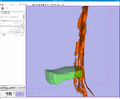
Double cylinder model used for the segmentation of the spine


Key Investigators
- Sylvain Jaume (MIT)
- Martin Loepprich (University of Heidelberg)
- Steve Pieper (BWH)
- Ron Kikinis (BWH)
- Polina Golland (MIT)
Objective
We are developing a Slicer module to segment the region within the thecal sac in MRI images of the spine. Our objective is to provide a segmentation and visualization tool to improve the treatment of disc herniation. The structures of interests are the cerebro-spinal fluid (CSF), the discs, the vertebrae and the spinal nerves. The main challenge is to perform the segmentation in a fully automated way.
Approach, Plan
Our plan for the project week is to integrate our code into Slicer 3.5. Our code analyzes the intensity profile of different regions in the MRI and automatically defines the optimum region for the CSF.
Progress
The algorithm will be implemented using ITK and VTK.
Screeshots
| [[Image:PW-SLC2010.png|Projects List |


References
- Segmentation using Slicer 3.5, EMSegment module