Difference between revisions of "Collaboration:CO-ME"
| (3 intermediate revisions by the same user not shown) | |||
| Line 1: | Line 1: | ||
Back to [[NA-MIC_External_Collaborations#International_Collaborations|NA-MIC External Collaborations]] | Back to [[NA-MIC_External_Collaborations#International_Collaborations|NA-MIC External Collaborations]] | ||
__NOTOC__ | __NOTOC__ | ||
| + | |||
| + | |||
| + | |||
| + | |||
| + | =NA-MIC Collaboration with Co-Me on Image-Guided Brain Tumor Surgery= | ||
| + | ==Key Personnel== | ||
| + | *Co-Me: Gabor Szekely | ||
| + | *NA-MIC: Ron Kikinis | ||
| + | |||
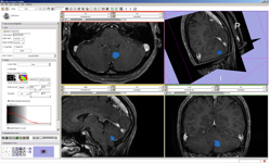
| + | [[Image:Collaboration ETH Come Image 1.png |thumb|left|300px|A pre-operative MRI from the USZ Neurosurgery viewed using 3D Slicer]] | ||
| + | |||
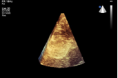
| + | [[Image:Collaboration ETH Come Image 2.png |thumb|right|300px|An intra-operative low-frequency 3D US image)]] | ||
| + | |||
| + | The National Centre of Competence in Research (NCCR) [http://co-me.ch/ Co-Me] is a network of leading clinics and engineering sites in Switzerland with strong links to industry and international partners. The leading house is the Swiss Federal Institute of Technology Zurich. The aim of the NCCR Co-Me is to take advantage of the computer technologies in order to improve patient care. | ||
| + | |||
| + | The goal of the collaboration is to exchange expertise in Neuronavigation for Brain Tumour Surgery. Specifically, we aim at providing computer assistance for supporting surgical decisions regarding the resection of tissue in an advanced phase of brain tumor surgery. The establishment of proper correspondences between pre- and intra-operatively acquired images by visual inspection is a significant challenge at this stage of the intervention and computer supported matching of those images can greatly contribute to performing the resection to a possibly full extent with maximal preservation of brain function. | ||
| + | |||
| + | Our core target is to create a neurosurgical navigation tool, enabling the fusion of pre-operative MRI with intra-operative MR and Ultrasound images in spite of the very significant geometrical and topological changes in the anatomy caused by tissue retraction and resection. The developed methods should also be able to cope with the major differences in tissue appearance on MR and US images as well as with the very limited field of view provided by high-frequency US imaging. Further challenge is to fully preserve the regularization power of the applied elastic deformation techniques while allowing for discontinuities in the underlying mapping. | ||
| + | |||
| + | The envisioned tool will be developed under the 3D Slicer platform, fully utilizing its current functionality for the generation of pre-operative brain models including tissue segmentation and the identification of critical structures such as fiber tracts or eloquent areas. Visualization and interactive information exploration will also rely on the available tools. The new methods for image registration and fusion will be fully integrated into this framework, which will be coupled with commercial navigation systems in order to provide direct intra-operative guidance. | ||
Latest revision as of 01:06, 6 May 2010
Home < Collaboration:CO-MEBack to NA-MIC External Collaborations
NA-MIC Collaboration with Co-Me on Image-Guided Brain Tumor Surgery
Key Personnel
- Co-Me: Gabor Szekely
- NA-MIC: Ron Kikinis
The National Centre of Competence in Research (NCCR) Co-Me is a network of leading clinics and engineering sites in Switzerland with strong links to industry and international partners. The leading house is the Swiss Federal Institute of Technology Zurich. The aim of the NCCR Co-Me is to take advantage of the computer technologies in order to improve patient care.
The goal of the collaboration is to exchange expertise in Neuronavigation for Brain Tumour Surgery. Specifically, we aim at providing computer assistance for supporting surgical decisions regarding the resection of tissue in an advanced phase of brain tumor surgery. The establishment of proper correspondences between pre- and intra-operatively acquired images by visual inspection is a significant challenge at this stage of the intervention and computer supported matching of those images can greatly contribute to performing the resection to a possibly full extent with maximal preservation of brain function.
Our core target is to create a neurosurgical navigation tool, enabling the fusion of pre-operative MRI with intra-operative MR and Ultrasound images in spite of the very significant geometrical and topological changes in the anatomy caused by tissue retraction and resection. The developed methods should also be able to cope with the major differences in tissue appearance on MR and US images as well as with the very limited field of view provided by high-frequency US imaging. Further challenge is to fully preserve the regularization power of the applied elastic deformation techniques while allowing for discontinuities in the underlying mapping.
The envisioned tool will be developed under the 3D Slicer platform, fully utilizing its current functionality for the generation of pre-operative brain models including tissue segmentation and the identification of critical structures such as fiber tracts or eloquent areas. Visualization and interactive information exploration will also rely on the available tools. The new methods for image registration and fusion will be fully integrated into this framework, which will be coupled with commercial navigation systems in order to provide direct intra-operative guidance.

