Difference between revisions of "2010 Summer Project Week RegistrationEvaluation"
Jfishbaugh (talk | contribs) |
Jfishbaugh (talk | contribs) |
||
| (18 intermediate revisions by the same user not shown) | |||
| Line 2: | Line 2: | ||
<gallery> | <gallery> | ||
Image:PW-MIT2010.png|[[2010_Summer_Project_Week#Projects|Projects List]] | Image:PW-MIT2010.png|[[2010_Summer_Project_Week#Projects|Projects List]] | ||
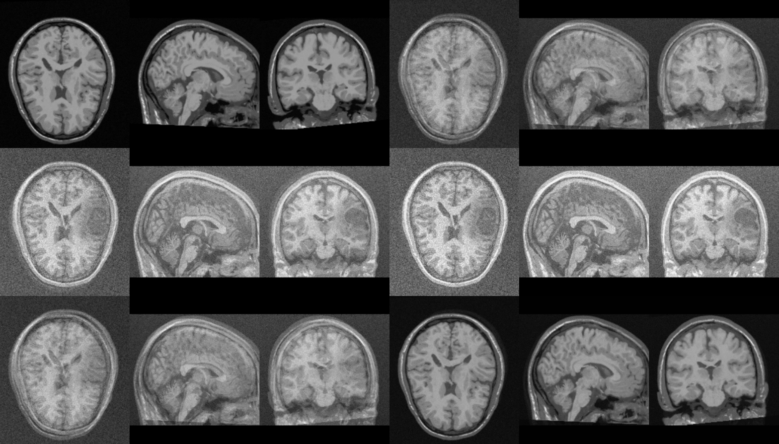
| + | Image:tumor_data.png | Data for experiment 1: Synthetic MRI with tumor | ||
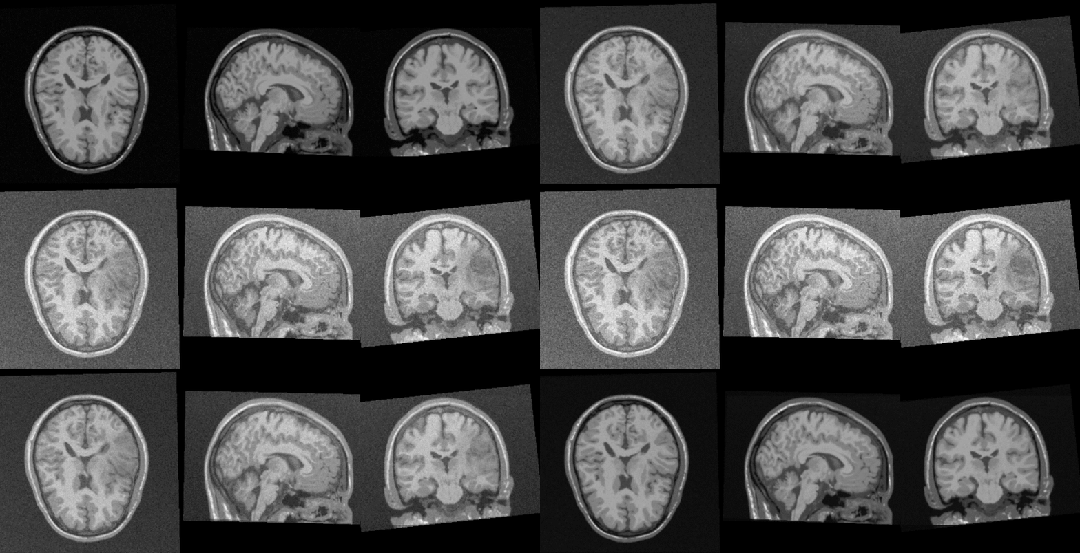
Image:TBI_smri.png | Data for experiment 2: Five modalities of sMRI for co-registration | Image:TBI_smri.png | Data for experiment 2: Five modalities of sMRI for co-registration | ||
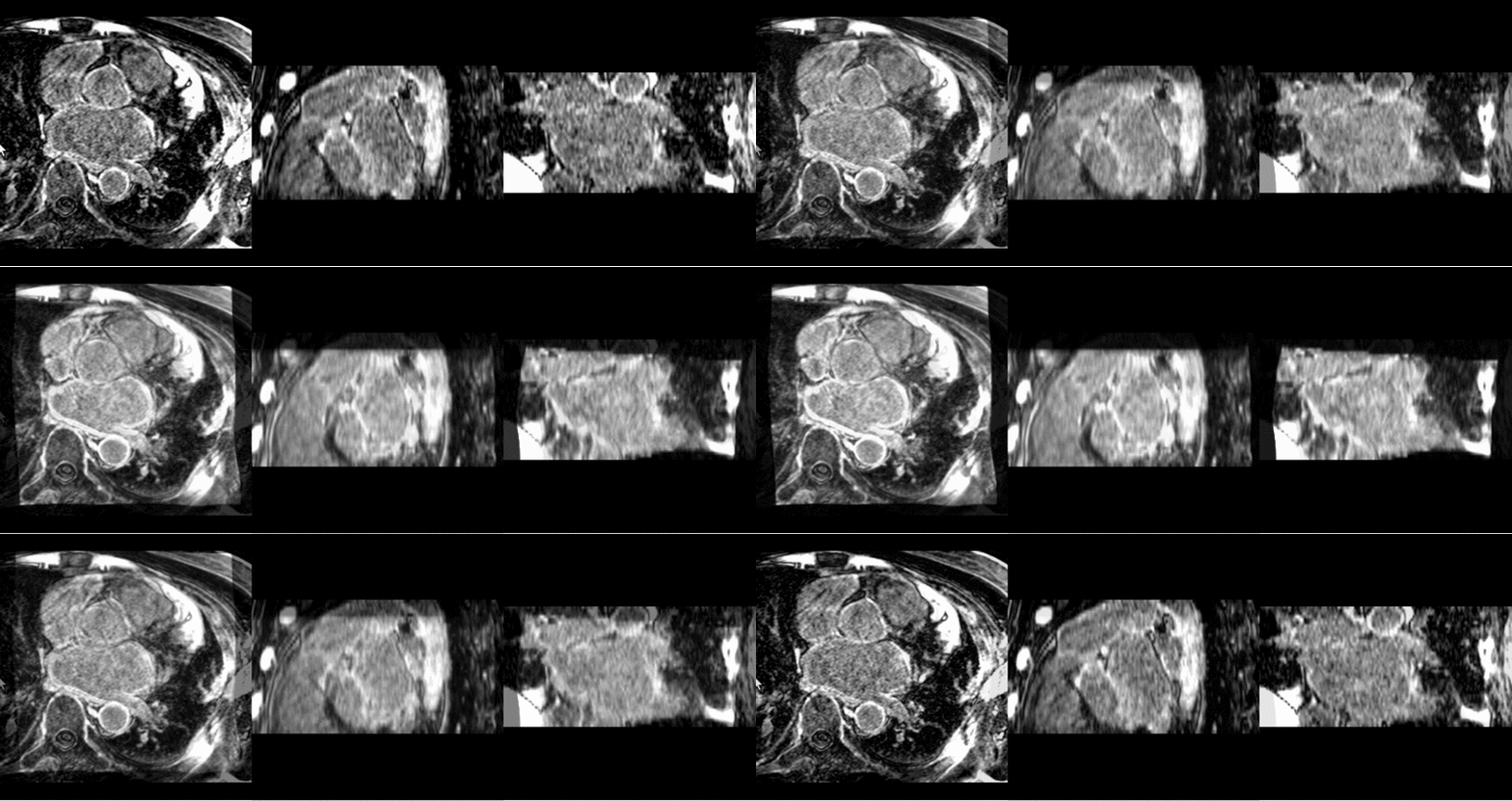
Image:afib_pre_vs_post.png | Data for experiment 3: Before and after ablation procedure | Image:afib_pre_vs_post.png | Data for experiment 3: Before and after ablation procedure | ||
| Line 35: | Line 36: | ||
This experiment consists of multi-modal MRI as well as DTI data from an individual with a severe head injury. We have 5 different modalities of structural MRI: axial TSE, axial GRE bleed, axial FLAIR, MP RAGE post-contrast, and SWI. We also have DTI data consisting of 20 directions. We would like to register the DTI data with the co-registered sMRI in order to view fiber tracts relative to the anatomy and pathology. | This experiment consists of multi-modal MRI as well as DTI data from an individual with a severe head injury. We have 5 different modalities of structural MRI: axial TSE, axial GRE bleed, axial FLAIR, MP RAGE post-contrast, and SWI. We also have DTI data consisting of 20 directions. We would like to register the DTI data with the co-registered sMRI in order to view fiber tracts relative to the anatomy and pathology. | ||
| − | This is useful experiment because the considerable contrast differences between the 5 modalities of structural MRI, as well as the different grid and pixel dimensions, provide an interesting multi-modal registration task. Furthermore, the non-linear nature of aligning DTI and sMRI | + | This is useful experiment because the considerable contrast differences between the 5 modalities of structural MRI, as well as the different grid and pixel dimensions, provide an interesting multi-modal registration task. Furthermore, the non-linear nature of aligning DTI and sMRI presents a challenging registration experiment. |
'''Experiment 3: Registration of Atrial Fibrillation Pre and Post Treatment''' | '''Experiment 3: Registration of Atrial Fibrillation Pre and Post Treatment''' | ||
| + | For this experiment, we are interested in registering structural MRI of the chest before and after ablation surgery. During the ablation procedure, a catheter is inserted to burn heart tissue in the atria demonstrating abnormal electrical activity. The ablation creates scar tissue, which does not conduct electricity, allowing the heart to return to a normal beating pattern. As a result of the procedure, the atrial wall shows a vastly different contrast than the scan taken before surgery. In addition, there are geometric differences between structures present in the two images. For these reasons, this is a challenging non-linear registration problem. | ||
</div> | </div> | ||
| Line 45: | Line 47: | ||
<h3>Progress</h3> | <h3>Progress</h3> | ||
| + | |||
| + | Good preliminary results have been obtained for all experiments. For these experiments, where the focus was on accuracy rather than speed of results or robustness across a population, we can conclude that the registration tools available in Slicer are of high quality. | ||
| + | |||
| + | Work needed to be completed: | ||
| + | |||
| + | * Written description of experiments | ||
| + | ** Parameters used | ||
| + | ** Command line calls to replicate results | ||
| + | * Analysis of results | ||
| + | **On synthetic example, comparison to known affine transformation | ||
| + | **Correlation, squared intensity error | ||
| + | * Further parameter exploration | ||
</div> | </div> | ||
| + | |||
| + | <div style="width: 97%; float: left; padding-right: 3%;"> | ||
| + | |||
| + | <h3>Results</h3> | ||
| + | |||
| + | '''Experiment 1: Synthetic Example''' | ||
| + | |||
| + | [[Image:synth_unreg.gif|thumb|center|800px|Data before registration.]] | ||
| + | |||
| + | [[Image:tumor_to_norm_notmasked.gif|thumb|center|800px|Affine registration without masking.]] | ||
| + | |||
| + | [[Image:tumor_to_norm_masked.gif|thumb|center|800px|Affine registration with masking.]] | ||
| + | |||
| + | [[Image:areg_tumor_to_norm.gif|thumb|center|800px|Affine registration using IRTK.]] | ||
| + | |||
| + | '''Experiment 2: Co-registration of Subject Specific Multi-modal MRI and DTI''' | ||
| + | |||
| + | [[Image:b0_unreg.gif|thumb|center|800px|Data before registration.]] | ||
| + | |||
| + | [[Image:b0_affine_reg.gif|thumb|center|800px|After affine registration.]] | ||
| + | |||
| + | [[Image:b0_bspline_reg.gif|thumb|center|800px|After BSpline registration.]] | ||
| + | |||
| + | [[Image:b0_to_tse_demons.gif|thumb|center|800px|After unconstrained diffeomorphic demons.]] | ||
| + | |||
| + | '''Experiment 3: Registration of Atrial Fibrillation Pre and Post Treatment''' | ||
| + | |||
| + | [[Image:post_to_pre_affine2.gif|thumb|center|800px|After affine registration.]] | ||
| + | |||
| + | [[Image:post_to_pre_bspline.gif|thumb|center|800px|After BSpline registration.]] | ||
| + | |||
| + | [[Image:post_to_pre_demons.gif|thumb|center|800px|After unconstrained diffeomorphic demons.]] | ||
| + | |||
| + | |||
| + | |||
| + | </div> | ||
| + | |||
</div> | </div> | ||
Latest revision as of 14:05, 25 June 2010
Home < 2010 Summer Project Week RegistrationEvaluationKey Investigators
- University of Utah: James Fishbaugh, Guido Gerig
- BWH: Dominik Meier
Objective
Recently, effort has been made in improving registration in Slicer. This includes the development and improvement of the RegisterImages module along with corresponding documentation from the use case library . The work on RegisterImages aimed at making registration easier, faster, and more accurate, while the use case library acts as a starting point and tutorial for users with a particular type of registration problem.
In order to continue improving registration, we propose a project to evaluate the current state of non-linear registration in Slicer. The results from this study will likely highlight areas of both strength and weakness in registration currently available in Slicer.
Approach, Plan
We propose evaluating registration in Slicer by focusing on three experiments:
Experiment 1: Synthetic Example
This experiment consists of registration between a baseline MRI and one with a synthetic tumor inserted via TumorSim. Furthermore, we will deform the baseline image with a known deformation field. We can evaluate both the transformed image for accuracy as well as the deformation field computed during registration. An interesting extension is to investigate how the size of the tumor or strength of the deformation affects registration accuracy.
Experiment 2: Co-registration of Subject Specific Multi-modal MRI and DTI
This experiment consists of multi-modal MRI as well as DTI data from an individual with a severe head injury. We have 5 different modalities of structural MRI: axial TSE, axial GRE bleed, axial FLAIR, MP RAGE post-contrast, and SWI. We also have DTI data consisting of 20 directions. We would like to register the DTI data with the co-registered sMRI in order to view fiber tracts relative to the anatomy and pathology.
This is useful experiment because the considerable contrast differences between the 5 modalities of structural MRI, as well as the different grid and pixel dimensions, provide an interesting multi-modal registration task. Furthermore, the non-linear nature of aligning DTI and sMRI presents a challenging registration experiment.
Experiment 3: Registration of Atrial Fibrillation Pre and Post Treatment
For this experiment, we are interested in registering structural MRI of the chest before and after ablation surgery. During the ablation procedure, a catheter is inserted to burn heart tissue in the atria demonstrating abnormal electrical activity. The ablation creates scar tissue, which does not conduct electricity, allowing the heart to return to a normal beating pattern. As a result of the procedure, the atrial wall shows a vastly different contrast than the scan taken before surgery. In addition, there are geometric differences between structures present in the two images. For these reasons, this is a challenging non-linear registration problem.
Progress
Good preliminary results have been obtained for all experiments. For these experiments, where the focus was on accuracy rather than speed of results or robustness across a population, we can conclude that the registration tools available in Slicer are of high quality.
Work needed to be completed:
- Written description of experiments
- Parameters used
- Command line calls to replicate results
- Analysis of results
- On synthetic example, comparison to known affine transformation
- Correlation, squared intensity error
- Further parameter exploration
Results
Experiment 1: Synthetic Example
Experiment 2: Co-registration of Subject Specific Multi-modal MRI and DTI
Experiment 3: Registration of Atrial Fibrillation Pre and Post Treatment


