Difference between revisions of "Projects:PathologyAnalysis"
| Line 20: | Line 20: | ||
</center> | </center> | ||
| − | |||
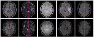
| − | [[File:Co-registration_result_No3.png]] | + | [[File:Co-registration_result_No3.png|thumb|400px|Result of coregistration]] |
Revision as of 14:32, 23 March 2011
Home < Projects:PathologyAnalysisBack to Utah 2 Algorithms
Analysis of Brain Images with Pathological Changes
Description
Traumatic brain injury (TBI) occurs when an external force traumatically injures the brain. TBI is a major cause of death and disability worldwide, especially in children and young adults. TBI affects 1.4 million Americans annually. The UCLA medical school has been working on this topic for years.
On anatomical MRI scans, to quantitatively analyze the cortical thickness, white matter changes, we need to have a good segmentation on TBI images. However, for TBI data, standard automated image analysis methods are not robust with respect to the TBI-related changes in image contrast, changes in brain shape, cranial fractures, white matter fiber alterations, and other signatures of head injury.
We are working on an extension of ABC for TBI datasets with the clinical goal to investigate alterations in cortical thickness, subsequent ventricular, and white matter changes in patients with TBI.
Key Investigators
- Utah: Bo Wang, Marcel Prastawa, Guido Gerig
- UCLA: Jack Van Horn, Andrei Irimia, Micah Chambers

