Difference between revisions of "DTI Tractography Challenge Tract"
| (3 intermediate revisions by the same user not shown) | |||
| Line 1: | Line 1: | ||
| − | + | '''Anatomical definition of the corticospinal tract''' <br> | |
| − | |||
| − | |||
| + | The corticospinal tract is a large bundle of about 1 million fibers that arise from the cerebral cortex, converge in the subcortical white matter (corona radiata) and course through the posterior limb of the internal capsule, the cerebral peduncle of the midbrain, the ventral pons (basis pontis), the ventral surface of the medulla, decussate in the lower medulla (pyramidal decussation), and terminate in the spinal cord. | ||
| + | The corticospinal tract contains fibers from the motor and premotor cortices (Broadmann areas 4 and 6), the primary somatosensory cortex (Brodmann areas 3,1, and 2), and from the superior parietal lobule (areas 5 and 7). It projects from the lateral medial cortex associated with the motor homunculus. | ||
| − | + | {| border="1" cellpadding="5" cellspacing="0" align="center" | |
| − | + | | colspan="3" align="center" | | |
| − | + | {| border="0" | |
| − | + | |+|- | |
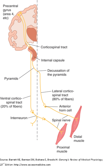
| − | The | + | | align="center" width="300px" |[[Image:Corticospinaltract.bmp|350px|thumb|Corticospinal tract. Source: Barrett KE, Barman SM, Boitano S, Brooks H. Ganong's Review of Medical Physiology, 23rd Edition. http://www.accessmedicine.com]] |
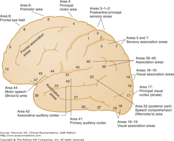
| + | | align="center" width="300px" |[[File:BrodmannAreas.bmp |thumb|350px|right|Lateral aspect of the cerebrum. The cortical areas are shown according to Brodmann with functional localizations. Source: Waxman SG. Clinical Neuroanatomy,26th Edition. http://www.accessmedicine.com]] | ||
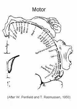
| + | | align="center" width="300px" |[[File:MotorHomunculus.bmp|thumb|250px|right|Motor Homunculus. Source: Penfield W. and Rasmussen T. The Cerebral Cortex of Man, New York, Macmillan, 1950.]] | ||
| + | |- | ||
| + | |} | ||
| + | |} | ||
Back to [[Events:_DTI_Tractography_Challenge_MICCAI_2011 | MICCAI 2011 DTI Tractography Challenge for Neurosurgical Planning]] | Back to [[Events:_DTI_Tractography_Challenge_MICCAI_2011 | MICCAI 2011 DTI Tractography Challenge for Neurosurgical Planning]] | ||
Latest revision as of 04:42, 30 March 2011
Home < DTI Tractography Challenge TractAnatomical definition of the corticospinal tract
The corticospinal tract is a large bundle of about 1 million fibers that arise from the cerebral cortex, converge in the subcortical white matter (corona radiata) and course through the posterior limb of the internal capsule, the cerebral peduncle of the midbrain, the ventral pons (basis pontis), the ventral surface of the medulla, decussate in the lower medulla (pyramidal decussation), and terminate in the spinal cord.
The corticospinal tract contains fibers from the motor and premotor cortices (Broadmann areas 4 and 6), the primary somatosensory cortex (Brodmann areas 3,1, and 2), and from the superior parietal lobule (areas 5 and 7). It projects from the lateral medial cortex associated with the motor homunculus.
| |||||
Back to MICCAI 2011 DTI Tractography Challenge for Neurosurgical Planning


