Difference between revisions of "Non-rigid MR-CT Image Registration"
| Line 1: | Line 1: | ||
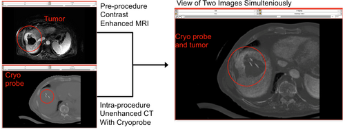
| − | [[Image:nonrigid.png |NON-RIGID MR-CT IMAGE REGISTRATION: This tutorial demonstrates how to perform MR-CT and CT-CT non-rigid registrations. In a cryoablation of liver case, we can see a tumor on pre-operated MRI. However, on intra-operated CT image, the tumor can not be seen though cryoproves can be seen. By using non-rigid MR-CT image registration, we can check the distance between the cryoprobes and tumor as shown the figure. |500px|thumb|right]] | + | [[Image:nonrigid.png |NON-RIGID MR-CT IMAGE REGISTRATION: This tutorial demonstrates how to perform MR-CT and CT-CT non-rigid registrations. In a CT-guided cryoablation of liver case, we can see a tumor on pre-operated MRI. However, on intra-operated CT image, the tumor can not be seen though cryoproves can be seen. By using non-rigid MR-CT image registration, we can check the distance between the cryoprobes and tumor as shown the figure. |500px|thumb|right]] |
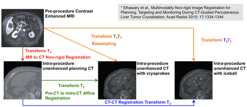
[[Image:nonrigid-cryo.png |PROCESS OF IMAGE REGISTRATION IN CRYOABLATION OF LIVER CASE: Registration process between MR and CT images in cryoablation of a liver case is shown. (1) A transformation matrix T1 was used to deform the pre-procedure contrast enhanced MR image on to the planning CT image. (2) T2 was used to deform the planning CT image on to the intra procedure CT image with cryoprobes. (3) The matrix T2 was combined with T1 to deform the MR image onto the CT image with cryoprobe. |500px|thumb|right]] | [[Image:nonrigid-cryo.png |PROCESS OF IMAGE REGISTRATION IN CRYOABLATION OF LIVER CASE: Registration process between MR and CT images in cryoablation of a liver case is shown. (1) A transformation matrix T1 was used to deform the pre-procedure contrast enhanced MR image on to the planning CT image. (2) T2 was used to deform the planning CT image on to the intra procedure CT image with cryoprobes. (3) The matrix T2 was combined with T1 to deform the MR image onto the CT image with cryoprobe. |500px|thumb|right]] | ||
Revision as of 01:23, 4 June 2011
Home < Non-rigid MR-CT Image Registration

Contents
Non-rigid MR-CT Image Registration Tutorial
Overview
This tutorial demonstrates how to perform MR-CT and Ct-CT non-rigid image registrations. The case study is CT-guided liver tumor cryoablation. In this tutorial, the three steps are performed as shown the figure on right side.
- (1) In MR-planning CT image registration, we can obtain the deformed MR image and the Bspline transformation matrix T1 by using pre-procedure contrast enhanced MRI and mask image.
- (2) In plannning CT-intraprocedure CT image registration, we can obtain the deformed planning CT image and the Bspline transformation matrix T2.
- (3) In MR-intraprocedure CT image registration, we can obtain the deformed MR image by using T1 and T2 BSpline transformation matrices.
For non-rigid registration, BRAINSFitIGT and BRAINSResamle modules are used. In this tutorial, we will show that 3D Slicer with BRAINSFitIGT module allows performing non-rigid image registration and BRAINSResample module allows performing non-rigid image deformation using Bspline transform matrix. We also shows in cryoablation of liver case, the distance between cryoprobe on CT image and tumor on MR image and the size of ice ball for the tumor can be confirmed easily by using the non-rigid MR-CT image registration.
Tutorials
Non-rigid MR-CT Image Registration Module tutorial : to perform MR-CT and CT-CT image registration [ppt] [pdf]
Tutorial materials
Material data:
People
Atsushi Yamada, Ph.D. (Research Associate, Brigham and Women's Hospital and Harvard Medical School)
Dominik S. Meier, Ph.D. (Assistant Professor, Brigham and Women's Hospital and Harvard Medical School)
Nobuhiko Hata, Ph.D. (Associate Professor, Brigham and Women's Hospital and Harvard Medical School)