Difference between revisions of "Collaboration:Marburg"
| Line 3: | Line 3: | ||
| + | [[image:Marburg-Logo-2011.png|right|thumb|300px]] | ||
=GBM Segmentation: Glioblastoma multiforme Segmentation under 3D Slicer for Neurosurgery= | =GBM Segmentation: Glioblastoma multiforme Segmentation under 3D Slicer for Neurosurgery= | ||
Revision as of 18:49, 26 July 2011
Home < Collaboration:MarburgBack to NA-MIC External Collaborations
Contents
GBM Segmentation: Glioblastoma multiforme Segmentation under 3D Slicer for Neurosurgery
Abstract
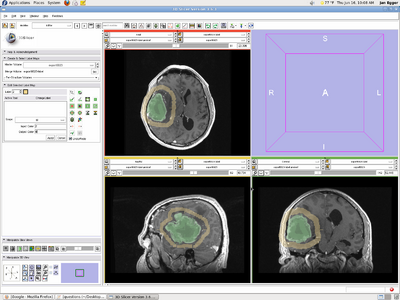
This project aims the evaluation of the (semi-)automated segmentation tools under the medical platform 3D Slicer. Thereby, the project focuses on World Health Organization (WHO) grad IV tumors (Glioblastoma multiforme) in Magnetic Resonance Imaging (MRI) datasets from the clinical routine. For evaluation of the methods, neurological surgeons with several years of experience in treatment of tumors perform manual slice by slice segmentation of dozens of WHO grade IV gliomas. Additionally, inter- and intra-physician segmentations have to be performed to provide a quality measure for the (semi-)automated segmentation tools. After segmentation, surface meshes – that consists of a set of triangles – of the segmented lesions have to be generated, voxelized and then the volume is calculated to enable a comparison via the Dice Similarity Coefficient (DSC). The Dice Similarity Coefficient is the relative volume overlap between A and R, where A and R are the binary masks from the automatic (A) and the reference (R) segmentation. V(·) is the volume (in cm3) of voxels inside the binary mask, by means of counting the number of voxels, then multiplying with the voxel size.
Key Personnel
- NA-MIC, NCIGT: Andriy Fedorov, Tina Kapur, Ron Kikinis, Rivka Colen, Alexandra Golby
- University Hospital of Marburg, Germany: Jan Egger, Bernd Freisleben, Christopher Nimsky
Projects
- Perform manual slice by slice segmentations of Glioblastoma multiforme (GBM) in Magnetic Resonance Imaging (MRI) datasets from the clinical routine.
- Achieve a quality measure for inter-physician segmentations by comparison of manual slice by slice segmentations from several neurological surgeons.
- Accomplish intra-physician segmentations to demonstrate the reproducibility performing manual boundary extraction and therefore provide a quality measure for automatic segmentations.
- Evaluation of the segmentation results achieved with the tools from 3D Slicer by using the manual segmentations under consideration of the accomplished inter- and intra-physician quality measures.
Study Design
- We would like to evaluate the performance of the existing tools in Slicer according to the following metrics:
- user time needed to do the segmentation
- amount of the cues/inputs provided by the user to the software
- quality of the overlap with the manual segmentations
Proposed study workflow:
|
Next steps:
- Alex/Tina identify two neurosurgeons
- Jan select 5 training cases
- Andrey/Jan train neurosurgeons
- Jan select 10 evaluation cases
- Andrey/Jan observe neurosurgeons during segmentation (measure time, save results)
- Andrey calculate DSC
Publications
- J. Egger, C. Kappus, B. Freisleben, Ch. Nimsky, A Medical Software System for Volumetric Analysis of Cerebral Pathologies in Magnetic Resonance Imaging (MRI) Data“, Journal of Medical Systems, Springer, Mar. 2011.
- J. Egger, Dz. Zukic, M.H.A. Bauer, D. Kuhnt, B. Carl, B. Freisleben, A. Kolb, Ch. Nimsky, A Comparison of Two Human Brain Tumor Segmentation Methods for MRI Data, Proceedings of 6th Russian-Bavarian Conference on Bio-Medical Engineering, State Technical University, Moscow, Russia, Nov. 2010.
- Dz. Zukic, J. Egger, M.H.A. Bauer, D. Kuhnt, B. Carl, B. Freisleben, A. Kolb, Ch. Nimsky, Glioblastoma Multiforme Segmentation in MRI Data with a Balloon Inflation Approach, Proceedings of 6th Russian-Bavarian Conference on Bio-Medical Engineering, State Technical University, Moscow, Russia, Nov. 2010.
- J. Egger, M. H. A. Bauer, D. Kuhnt, C. Kappus, B. Carl, B. Freisleben, Ch. Nimsky, Evaluation of a Novel Approach for Automatic Volume Determination of Glioblastomas Based on Several Manual Expert Segmentations, Proceedings of 44. Jahrestagung der DGBMT, Rostock, Germany, Oct. 2010.
- J. Egger, M. H. A. Bauer, D. Kuhnt, B. Freisleben, Ch. Nimsky, Min-Cut-Segmentation of WHO Grade IV Gliomas Evaluated against Manual Segmentation, XIX Congress of the European Society for Stereotactic and Functional Neurosurgery, Athens, Greece, Sep. 2010.
- J. Egger, M. H. A. Bauer, D. Kuhnt, B. Carl, C. Kappus, B. Freisleben, Ch. Nimsky, Nugget-Cut: A Segmentation Scheme for Spherically- and Elliptically-Shaped 3D Objects, 32nd Annual Symposium of the German Association for Pattern Recognition (DAGM), LNCS 6376, pp. 383–392, Springer Press, Darmstadt, Germany, Sep. 2010.
- J. Egger, M. H. A. Bauer, D. Kuhnt, C. Kappus, B. Carl, B. Freisleben, Ch. Nimsky. A Flexible Semi-Automatic Approach for Glioblastoma multiforme Segmentation. Proceedings of International Biosignal Processing Conference, Charité, Berlin, Germany, Jul. 2010.
Resource Links
- 3D Slicer (http://www.slicer.org/)
- GrowCutSegmentation (http://www.slicer.org/slicerWiki/index.php/Modules:GrowCutSegmentation-Documentation-3.6)
- Neurosurgery Department of Marburg (http://www.ukgm.de/ugm_2/deu/umr_nch/umr_nch_team.php?id=767)

