Difference between revisions of "DBP3:Utah:Reg Motiv"
From NAMIC Wiki
(Created page with '= Left Atrial Registration = * Targeting the LA wall and pulmonary veins') |
|||
| Line 1: | Line 1: | ||
= Left Atrial Registration = | = Left Atrial Registration = | ||
* Targeting the LA wall and pulmonary veins | * Targeting the LA wall and pulmonary veins | ||
| + | |||
| + | {| class="wikitable" | ||
| + | |- | ||
| + | ! Pre-ablation LGE-MRI Image | ||
| + | ! Post-ablation LGE-MRI Image | ||
| + | |- | ||
| + | | [[File:carma_ex_pre.png|thumb|center]] | ||
| + | | [[File:carma_ex_post.png|thumb|center]] | ||
| + | |- | ||
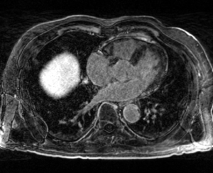
| + | | MRI image acquired approx. 15 minutes after contrast injection and prior to ablation. (The LA is the gray, horizontally oriented structure above the spine in the center of the image) | ||
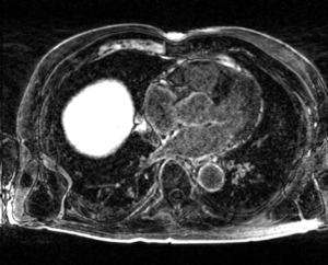
| + | | MRI image acquired approx. 15 minutes after contrast injection and after the ablation was performed. Sections of the LA wall which were ablated appear as hyper-enhanced pixels in the LA wall. | ||
| + | |- | ||
| + | ! MRA Image | ||
| + | ! Segmentation of LGE-MRI Image | ||
| + | |- | ||
| + | | [[File:carma_ex_mra.png|thumb|center]] | ||
| + | | [[File:carma_ex_pre_seg.png|thumb|center]] | ||
| + | |- | ||
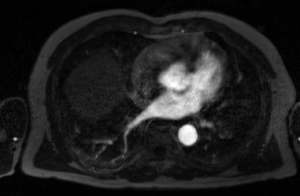
| + | | The MRA is acquired after contrast injection and before the LGE-MRI images above. This image type highlights the blood pool of the LA and some surrounding structures. | ||
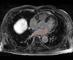
| + | | manual segmentation of the LA is performed on pre- and post-ablation LGE-MRI images by the CARMA team. Here the blue mask denotes the LA wall and the orange the blood pool. | ||
| + | |- | ||
| + | |} | ||
Revision as of 22:53, 6 January 2012
Home < DBP3:Utah:Reg MotivLeft Atrial Registration
- Targeting the LA wall and pulmonary veins
| Pre-ablation LGE-MRI Image | Post-ablation LGE-MRI Image |
|---|---|
| MRI image acquired approx. 15 minutes after contrast injection and prior to ablation. (The LA is the gray, horizontally oriented structure above the spine in the center of the image) | MRI image acquired approx. 15 minutes after contrast injection and after the ablation was performed. Sections of the LA wall which were ablated appear as hyper-enhanced pixels in the LA wall. |
| MRA Image | Segmentation of LGE-MRI Image |
| The MRA is acquired after contrast injection and before the LGE-MRI images above. This image type highlights the blood pool of the LA and some surrounding structures. | manual segmentation of the LA is performed on pre- and post-ablation LGE-MRI images by the CARMA team. Here the blue mask denotes the LA wall and the orange the blood pool. |


