Difference between revisions of "2012 Winter Project Week:TBIValidation"
From NAMIC Wiki
| (17 intermediate revisions by the same user not shown) | |||
| Line 3: | Line 3: | ||
Image:PW-SLC2012.png|[[2012_Winter_Project_Week#Projects|Projects List]] | Image:PW-SLC2012.png|[[2012_Winter_Project_Week#Projects|Projects List]] | ||
</gallery> | </gallery> | ||
| + | |||
| + | <center> | ||
| + | <gallery widths=270px heights=220px perrow=2 caption="Visualization and validation of segmentation "> | ||
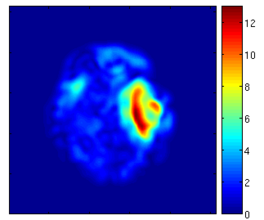
| + | File:DefVis DetJacobian TBISeg.png|Visualization of the deformation field via determinant of Jacobian | ||
| + | File:DefVis VecMag TBISeg.png|Visualization of the deformation field via vector magnitude | ||
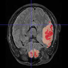
| + | File:Manual_seg_2D_X_view.jpg|Manual segmentation - ground truth | ||
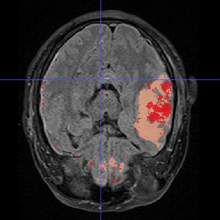
| + | File:Semiauto_seg_2D_X_view.jpg|Result of the segmentation algorithm | ||
| + | </gallery> | ||
| + | </center> | ||
| + | |||
==Key Investigators== | ==Key Investigators== | ||
| − | Marcel Prastawa, | + | |
| + | *Utah: Bo Wang, Marcel Prastawa, and Guido Gerig | ||
| + | *UCLA: Andrei Irimia, Micah Chambers, Jack Van Horn and Paul M. Vespa | ||
| + | *Kitware: Danielle Pace, Stephen Aylward | ||
<div style="margin: 20px;"> | <div style="margin: 20px;"> | ||
| Line 11: | Line 24: | ||
<h3>Objective</h3> | <h3>Objective</h3> | ||
| − | * | + | * Define manual segmentation protocol for MR images of TBI |
| − | * | + | * Validate the segmentation of the algorithm |
| − | * | + | * Visualize volume changes, deformation field |
</div> | </div> | ||
| Line 21: | Line 34: | ||
<h3>Approach, Plan</h3> | <h3>Approach, Plan</h3> | ||
Our plan for the project week: | Our plan for the project week: | ||
| − | * | + | * Work with collaborators to define manual segmentation protocol |
| − | * | + | * Compare the independent segmentation and joint segmentation |
| + | * Visualize volume changes and deformation field according to clinicians' requirements. | ||
</div> | </div> | ||
| Line 29: | Line 43: | ||
<h3>Progress</h3> | <h3>Progress</h3> | ||
| − | + | * Summarized the rule the clinician used to do manual segmentation | |
| + | * Compared the result of the algorithm with clinician's manual segmentation | ||
| + | * Explored different ways to visualize the volume changes/deformation field, we tried the following methods. | ||
| + | ** Jacobian determinant | ||
| + | ** Vector magnitude | ||
</div> | </div> | ||
Latest revision as of 15:44, 13 January 2012
Home < 2012 Winter Project Week:TBIValidation- Visualization and validation of segmentation
Key Investigators
- Utah: Bo Wang, Marcel Prastawa, and Guido Gerig
- UCLA: Andrei Irimia, Micah Chambers, Jack Van Horn and Paul M. Vespa
- Kitware: Danielle Pace, Stephen Aylward
Objective
- Define manual segmentation protocol for MR images of TBI
- Validate the segmentation of the algorithm
- Visualize volume changes, deformation field
Approach, Plan
Our plan for the project week:
- Work with collaborators to define manual segmentation protocol
- Compare the independent segmentation and joint segmentation
- Visualize volume changes and deformation field according to clinicians' requirements.
Progress
- Summarized the rule the clinician used to do manual segmentation
- Compared the result of the algorithm with clinician's manual segmentation
- Explored different ways to visualize the volume changes/deformation field, we tried the following methods.
- Jacobian determinant
- Vector magnitude
References
- Bo Wang, Marcel Prastawa, Andrei Irimia, Micah C. Chambers, Paul M. Vespa, John D. van Horn, Guido Gerig, A Patient-Specific Segmentation Framework for Longitudinal MR Images of Traumatic Brain Injury, SPIE Medical Imaging 2012.
- Andrei Irimia, Micah C. Chambers, Jeffry R. Alger, Maria Filippou, Marcel W. Prastawa, Bo Wang, David A. Hovda, Guido Gerig, Arthur W. Toga, Ron Kikinis, Paul M. Vespa, John D. van Horn (2011) Comparison of acute and chronic traumatic brain injury using semi-automatic multimodal segmentation of MR volumes. Journal of Neurotrauma


