Difference between revisions of "2013 Summer Project Week:Epilepsy Surgery"
From NAMIC Wiki
| Line 74: | Line 74: | ||
<br> | <br> | ||
[[File:tlNoblur.png]] | [[File:tlNoblur.png]] | ||
| − | [[File:histNoblur.png]] | + | [[File:histNoblur.png]] Without blurring |
[[File:tlBlurred.png]] | [[File:tlBlurred.png]] | ||
| − | [[File:histBlurred.png]] | + | [[File:histBlurred.png]] With blurring |
<br> | <br> | ||
Revision as of 05:34, 21 June 2013
Home < 2013 Summer Project Week:Epilepsy SurgeryKey Investigators
- USP - Luiz Murta
Objective
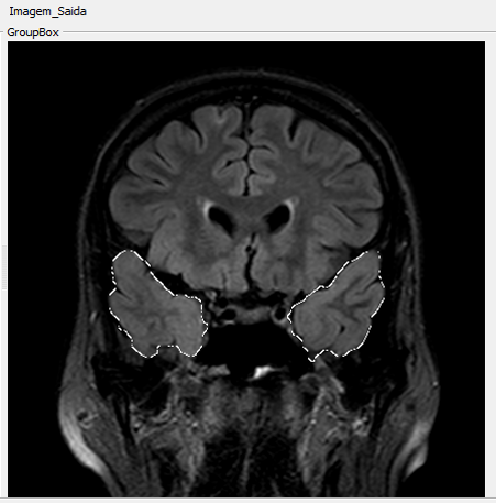
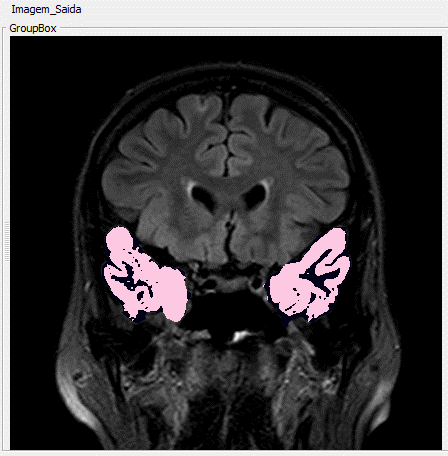
This project will investigate the presence and location of the epileptogenic focus in temporal lobe by analyzing patterns of texture in magnetic resonance imaging (MRI) after segmentation using anisotropic diffusion filters anomalous and geodesic active contour.
Purpose
Progress
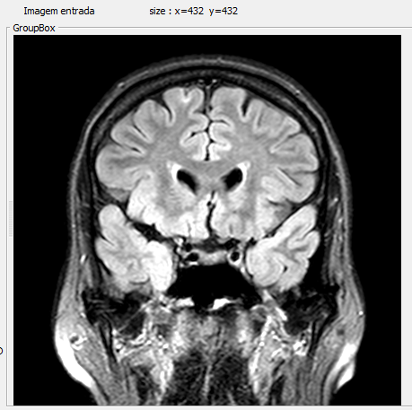
Examples:
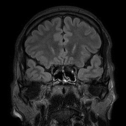
Normal MRI at mesial temporal lobe
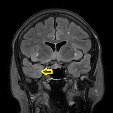
MRI containing blurring phenomena on right side as indicated by the yellow arrow
Results
Classifier
-- artificial neural network;
-- nearest neighbour;
-- and decision tree
-- 24 were extracted from the co-occurrence matrix using Haralick texture descriptors and
-- 8 were intensity statistics obtained from histogram.
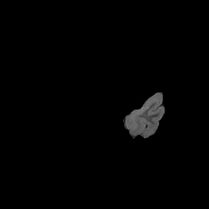
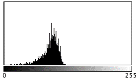 Without blurring
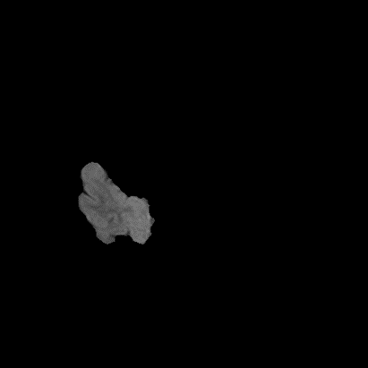
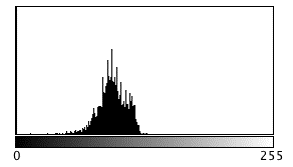 With blurring
Decision Tree
D_Hist_MeanIntensity <= 0.453
| D_Hist_stdDev <= 0.138: with_blurring (5.0)
| D_Hist_stdDev > 0.138
| | E_Hist_Kurtosis <= 0.497
| | | E_COOmeanHomogeneity_dist3 <= 0.425
| | | | D_COOmeanHomogeneity_dist2 <= 0.318: without_blurring (9.0/2.0)
| | | | D_COOmeanHomogeneity_dist2 > 0.318: with_blurring (5.0)
| | | E_COOmeanHomogeneity_dist3 > 0.425
| | | | E_COOmeanHomogeneity_dist3 <= 0.895: without_blurring (26.0/1.0)
| | | | E_COOmeanHomogeneity_dist3 > 0.895: with_blurring (3.0/1.0)
| | E_Hist_Kurtosis > 0.497: with_blurring (5.0)
D_Hist_MeanIntensity > 0.453
| E_Hist_Kurtosis <= 0.48: with_blurring (12.0)
| E_Hist_Kurtosis > 0.48
| | E_COOmeanHomogeneity_dist1 <= 0.432: with_blurring (3.0)
| | E_COOmeanHomogeneity_dist1 > 0.432: without_blurring (2.0)
Number of Leaves : 9
Conclusions
References
- Shaker, M. & Soltanian-Zadeh, H., 2008. Voxel-Based Morphometric Study of Brain Regions from Magnetic Resonance Images in Temporal Lobe Epilepsy. Image Analysis and Interpretation, 2008. SSIAI 2008. IEEE Southwest Symposium on, 209-212.

