Difference between revisions of "SPIE 2013 DTI Workshop"
(Created page with '{| class="wikitable" border="1" cellpadding="8" cellspacing="1" | colspan="2" style="width:100px" |Image:spie2012.gif |- | style="width:50%" |Image:NAMIC.jpg | style="…') |
|||
| (31 intermediate revisions by 3 users not shown) | |||
| Line 11: | Line 11: | ||
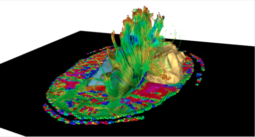
The development of Diffusion Tensor Magnetic Resonance Imaging (DT-MRI) has opened up the possibility of studying the complex organization of the brain's white matter in-vivo. By measuring the diffusion of water molecules in tissues, the technique gives insights into the structure and orientation of major white matter pathways, and DT-MRI findings have the potential to play a critical role in the extraction of meaningful information for diagnosis, prognosis and following of treatment response. | The development of Diffusion Tensor Magnetic Resonance Imaging (DT-MRI) has opened up the possibility of studying the complex organization of the brain's white matter in-vivo. By measuring the diffusion of water molecules in tissues, the technique gives insights into the structure and orientation of major white matter pathways, and DT-MRI findings have the potential to play a critical role in the extraction of meaningful information for diagnosis, prognosis and following of treatment response. | ||
| − | The course will guide participants through the fundamental aspects of DT-MRI data analysis, as well as the challenges of transferring cutting-edge DT-MRI techniques to clinical routine. The format will include a series of hands-on sessions with the participants running DT-MRI analysis on their own laptops, to provide a practical experience of extracting useful clinical information from Diffusion MR images. The hands-on sessions will use DT-MRI tools from the NA-MIC toolkit, which include the 3DSlicer software, an open-source platform for medical image processing and 3D visualization used in biomedical and clinical research. Participants will be guided through an integrated workflow for exploring the brain white matter in a series of datasets that will be provided as part of the course. This event is part of the on-going effort of the NIH-funded National Alliance for Medical Image Computing (NA-MIC) to transfer the latest advances in biomedical image analysis to the scientific and clinical community. | + | The [http://spie.org/app/program/index.cfm?fuseaction=COURSE&export_id=x12534&ID=x12172&redir=x12172.xml&course_id=E2011888&event_id=896166&programtrack_id=37 SPIE 2013 course] will guide participants through the fundamental aspects of DT-MRI data analysis, as well as the challenges of transferring cutting-edge DT-MRI techniques to clinical routine. The format will include a series of hands-on sessions with the participants running DT-MRI analysis on their own laptops, to provide a practical experience of extracting useful clinical information from Diffusion MR images. The hands-on sessions will use DT-MRI tools from the NA-MIC toolkit, which include the 3DSlicer software, an open-source platform for medical image processing and 3D visualization used in biomedical and clinical research. Participants will be guided through an integrated workflow for exploring the brain white matter in a series of datasets that will be provided as part of the course. This event is part of the on-going effort of the NIH-funded National Alliance for Medical Image Computing (NA-MIC) to transfer the latest advances in biomedical image analysis to the scientific and clinical community. |
| + | |||
| + | ==Faculty == | ||
| + | *Sonia Pujol, Ph.D., Surgical Planning Laboratory, Brigham and Women’s Hospital, Harvard Medical School | ||
| + | *Guido Gerig, Ph.D., The Scientific Computing and Imaging Institute, University of Utah | ||
| + | *Martin Styner, Ph.D.,Neuro Image Research and Analysis Laboratory, University of North Carolina | ||
| + | |||
== Date and Location== | == Date and Location== | ||
| + | * Saturday February 9, 2013 | ||
[http://spie.org/x12166.xml SPIE Medical Imaging 2013, Orlando, Florida]. | [http://spie.org/x12166.xml SPIE Medical Imaging 2013, Orlando, Florida]. | ||
| − | |||
== Registration== | == Registration== | ||
| Line 33: | Line 39: | ||
== Intended Audience == | == Intended Audience == | ||
Scientists, engineers, and clinical researchers who are interested in learning how to use Diffusion Tensor MR Imaging for mapping the white matter of the human brain in health and disease. This course does not need any prior knwoledge, but it can be combined with the course [http://spie.org/app/program/index.cfm?fuseaction=COURSE&export_id=x12534&ID=x12172&redir=x12172.xml&course_id=E0982442&event_id=896165 SC 1063: Diffusion Imaging]. | Scientists, engineers, and clinical researchers who are interested in learning how to use Diffusion Tensor MR Imaging for mapping the white matter of the human brain in health and disease. This course does not need any prior knwoledge, but it can be combined with the course [http://spie.org/app/program/index.cfm?fuseaction=COURSE&export_id=x12534&ID=x12172&redir=x12172.xml&course_id=E0982442&event_id=896165 SC 1063: Diffusion Imaging]. | ||
| + | |||
| + | == Tentative Agenda == | ||
| + | |||
| + | *12:30-1:30 pm Pre-workshop installation of software and datasets: [[Media:Spie2013SC1065UNCSlicerTutorial.pdf | Slides Extension Installation (PDF)]] | ||
| + | *1:30-1:35 pm Introduction to the course (Sonia Pujol) | ||
| + | *1:35-2:00 pm Fundamentals of DTI analysis (Guido Gerig) | ||
| + | *2:00-2:10 pm Presentation of the hands-on DTI pipeline ** (Sonia Pujol) | ||
| + | *2:10-2:40 pm Dicom conversion and DWI Quality Control (Martin Styner): [[Media:2013-SPIE-1-DICOMToNRRDConversionTutorial.pptx| Slides Dicom Conversion(PPTX)]] / [[Media:2013-SPIE-2-DTIQC.pptx| Slides DTIPrep QC (PPTX) ]] | ||
| + | *2:40-3:10 pm DTI (pairwise) registration for clinical studies (Martin Styner): [[Media:2013-SPIE-3-DTI-Reg-Tutorial.pptx | Slides DTI-Reg (PPTX)]] | ||
| + | |||
| + | *3:10-3:25 pm Coffee Break | ||
| + | |||
| + | *3:25-3:50 pm DTI unbiased atlas building for population studies (Martin Styner) [[Media:2013-SPIE-4-DTIAtlasBuilder_Tutorial.pptx | Slides DTIAtlasBuilder (PPTX)]] | ||
| + | *3:50-4:50 pm [[media:DiffusionTensorImaging SPIE2013 SoniaPujol.pdf | From DWI images to fiber tracts (Sonia Pujol) (Tensor Estimation, Glyphs, Scalar Diffusion Measurements, Fiber Tracking) ]] | ||
| + | *4:50-5:15 pm Towards validation of DTI Tractography (Sonia Pujol) | ||
| + | *5:15-5:30 pm Conclusion and Questions from the audience | ||
| + | |||
| + | * Additional slides: | ||
| + | ** FiberViewerLight tutorial ([[media:2012-SPIE-FiberViewerLightTutorial.pptx | slides from 2012]]) | ||
| + | ** DTI fiber profile processing and analysis via DTIFiberAtlasAnalyzer | ||
==Preparation for the hands-on sessions: Software and datasets == | ==Preparation for the hands-on sessions: Software and datasets == | ||
The workshop combines oral presentations and instructor-led hands-on sessions with the participants working on their own laptop computers. | The workshop combines oral presentations and instructor-led hands-on sessions with the participants working on their own laptop computers. | ||
| − | All participants are required to come with their own laptop computer and install the software and datasets prior to the event. A minimum of 2 GB of RAM (4 GB is better) and a graphic accelerator with 256 MB (512MB is better) of on-board graphic memory are required. The 3DSlicer version 4 software | + | All participants are required to come with their own laptop computer and install the software and datasets prior to the event. A minimum of 2 GB of RAM (4 GB is better) and a graphic accelerator with 256 MB (512MB is better) of on-board graphic memory are required. |
| + | |||
| + | *The 3DSlicer version 4.2.2.1 software will need to be downloaded from here: [http://download.slicer.org Slicer download page (version r21513)] | ||
| + | *Additional instructions on how to install the DTI processing extensions will be provided next week. | ||
| + | *The datasets to download will be posted a few days before the event on this website. | ||
| + | *In addition, time is allotted to install the data from USB sticks at the beginning of the workshop. | ||
The following OS are supported: MacOS X Lion, Windows 7 (VS 2008), Linux 64. | The following OS are supported: MacOS X Lion, Windows 7 (VS 2008), Linux 64. | ||
| Line 46: | Line 77: | ||
[http://www.surveymonkey.com/s/GZDXKXQ Click here to take the Slicer4 Training Survey] | [http://www.surveymonkey.com/s/GZDXKXQ Click here to take the Slicer4 Training Survey] | ||
| − | + | ==NA-MIC Presence at SPIE 2013 == | |
| + | * [http://spie.org/Documents/ConferencesExhibitions/MI2013-Final-lr.pdf DTI/Functional session, Sunday Feb.10, 2013 10:10 am -12:10 pm] Session Chair: Sonia Pujol, Ph.D. | ||
| + | * [http://spie.org/Documents/ConferencesExhibitions/MI2013-Final-lr.pdf Temporal and Motional Analysis, Sunday Feb.10, 2013, 3:30 pm -5:30 pm] Session Chair: Martin Styner, Ph.D. | ||
Back to [http://www.na-mic.org/Wiki/index.php/Events NA-MIC Events] | Back to [http://www.na-mic.org/Wiki/index.php/Events NA-MIC Events] | ||
Latest revision as of 14:06, 30 August 2013
Home < SPIE 2013 DTI Workshop
| |

|

|
Exploring Brain Connectivity in-vivo: from Theory to Practice - A hands-on analysis workshop on Diffusion MRI by the National Alliance for Medical Image Computing (NA-MIC)
Course Description:
The development of Diffusion Tensor Magnetic Resonance Imaging (DT-MRI) has opened up the possibility of studying the complex organization of the brain's white matter in-vivo. By measuring the diffusion of water molecules in tissues, the technique gives insights into the structure and orientation of major white matter pathways, and DT-MRI findings have the potential to play a critical role in the extraction of meaningful information for diagnosis, prognosis and following of treatment response. The SPIE 2013 course will guide participants through the fundamental aspects of DT-MRI data analysis, as well as the challenges of transferring cutting-edge DT-MRI techniques to clinical routine. The format will include a series of hands-on sessions with the participants running DT-MRI analysis on their own laptops, to provide a practical experience of extracting useful clinical information from Diffusion MR images. The hands-on sessions will use DT-MRI tools from the NA-MIC toolkit, which include the 3DSlicer software, an open-source platform for medical image processing and 3D visualization used in biomedical and clinical research. Participants will be guided through an integrated workflow for exploring the brain white matter in a series of datasets that will be provided as part of the course. This event is part of the on-going effort of the NIH-funded National Alliance for Medical Image Computing (NA-MIC) to transfer the latest advances in biomedical image analysis to the scientific and clinical community.
Faculty
- Sonia Pujol, Ph.D., Surgical Planning Laboratory, Brigham and Women’s Hospital, Harvard Medical School
- Guido Gerig, Ph.D., The Scientific Computing and Imaging Institute, University of Utah
- Martin Styner, Ph.D.,Neuro Image Research and Analysis Laboratory, University of North Carolina
Date and Location
- Saturday February 9, 2013
SPIE Medical Imaging 2013, Orlando, Florida.
Registration
To register for this course, please visit the SPIE 2013 conference website.
Learning Outcomes
This course will enable participants to:
- identify the different components of a DT-MRI fiber tract analysis pipeline
- perform DWI/DTI data quality control
- visualize 3D tensor fields and diffusion-derived maps
- generate 3D reconstructions of white matter tracts in a normal subject and pathological case
- extract and visualize DTI fiber tract profiles
- identify the current challenges inherent in using DT-MRI data in the clinics
For questions related to the workshop, please send an e-mail to Sonia Pujol (spujol at bwh.harvard.edu).
Intended Audience
Scientists, engineers, and clinical researchers who are interested in learning how to use Diffusion Tensor MR Imaging for mapping the white matter of the human brain in health and disease. This course does not need any prior knwoledge, but it can be combined with the course SC 1063: Diffusion Imaging.
Tentative Agenda
- 12:30-1:30 pm Pre-workshop installation of software and datasets: Slides Extension Installation (PDF)
- 1:30-1:35 pm Introduction to the course (Sonia Pujol)
- 1:35-2:00 pm Fundamentals of DTI analysis (Guido Gerig)
- 2:00-2:10 pm Presentation of the hands-on DTI pipeline ** (Sonia Pujol)
- 2:10-2:40 pm Dicom conversion and DWI Quality Control (Martin Styner): Slides Dicom Conversion(PPTX) / Slides DTIPrep QC (PPTX)
- 2:40-3:10 pm DTI (pairwise) registration for clinical studies (Martin Styner): Slides DTI-Reg (PPTX)
- 3:10-3:25 pm Coffee Break
- 3:25-3:50 pm DTI unbiased atlas building for population studies (Martin Styner) Slides DTIAtlasBuilder (PPTX)
- 3:50-4:50 pm From DWI images to fiber tracts (Sonia Pujol) (Tensor Estimation, Glyphs, Scalar Diffusion Measurements, Fiber Tracking)
- 4:50-5:15 pm Towards validation of DTI Tractography (Sonia Pujol)
- 5:15-5:30 pm Conclusion and Questions from the audience
- Additional slides:
- FiberViewerLight tutorial ( slides from 2012)
- DTI fiber profile processing and analysis via DTIFiberAtlasAnalyzer
Preparation for the hands-on sessions: Software and datasets
The workshop combines oral presentations and instructor-led hands-on sessions with the participants working on their own laptop computers. All participants are required to come with their own laptop computer and install the software and datasets prior to the event. A minimum of 2 GB of RAM (4 GB is better) and a graphic accelerator with 256 MB (512MB is better) of on-board graphic memory are required.
- The 3DSlicer version 4.2.2.1 software will need to be downloaded from here: Slicer download page (version r21513)
- Additional instructions on how to install the DTI processing extensions will be provided next week.
- The datasets to download will be posted a few days before the event on this website.
- In addition, time is allotted to install the data from USB sticks at the beginning of the workshop.
The following OS are supported: MacOS X Lion, Windows 7 (VS 2008), Linux 64.
Slicer Community
Participants are invited to join the Slicer user and Slicer developer mailing lists prior to the workshop. This is a place for the Slicer community to discuss questions and feature requests related to 3D Slicer.
Slicer4 Training Survey
Click here to take the Slicer4 Training Survey
NA-MIC Presence at SPIE 2013
- DTI/Functional session, Sunday Feb.10, 2013 10:10 am -12:10 pm Session Chair: Sonia Pujol, Ph.D.
- Temporal and Motional Analysis, Sunday Feb.10, 2013, 3:30 pm -5:30 pm Session Chair: Martin Styner, Ph.D.
Back to NA-MIC Events
