Difference between revisions of "Organ Detection"
From NAMIC Wiki
| (2 intermediate revisions by one other user not shown) | |||
| Line 5: | Line 5: | ||
==Key Investigators== | ==Key Investigators== | ||
| − | German Gonzalez, James Ross, Raul San Jose | + | German Gonzalez, James Ross, Raul San Jose (Brigham and Women's Hospital) |
==Project Description== | ==Project Description== | ||
| Line 22: | Line 22: | ||
<div style="width: 27%; float: left; padding-right: 3%;"> | <div style="width: 27%; float: left; padding-right: 3%;"> | ||
<h3>Progress</h3> | <h3>Progress</h3> | ||
| − | * | + | * Python prototype working for different chest structures like pulmonary artery, right ventricle, left ventricle. |
</div> | </div> | ||
</div> | </div> | ||
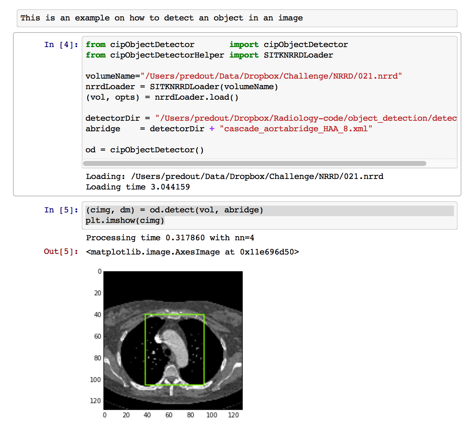
| + | [[File:DemoObjectDetection.png]] | ||
Latest revision as of 16:32, 9 January 2015
Home < Organ DetectionKey Investigators
German Gonzalez, James Ross, Raul San Jose (Brigham and Women's Hospital)
Project Description
Objective
- Develop an organ ROI detection tool for 2D slides to enable quick initialization and rapid
Approach, Plan
- Leverage OpenCV technology for object detection in images.
- Integration of OpenCV tools as a Slicer module to enable training and deployment of object detectors
- Discuss other alternatives:
- Jim recommends looking at the 2013 SPIE paper that used the histogram of oriented gradients.
Progress
- Python prototype working for different chest structures like pulmonary artery, right ventricle, left ventricle.

