Difference between revisions of "Project Week 25/Surgical Planning In Stereotaxy"
m (Fill progress and next steps section) |
m |
||
| (3 intermediate revisions by the same user not shown) | |||
| Line 6: | Line 6: | ||
==Key Investigators== | ==Key Investigators== | ||
<!-- Key Investigator bullet points --> | <!-- Key Investigator bullet points --> | ||
| + | * Fernando Pérez-García ([http://icm-institute.org/en/ Brain & Spine Institute], Paris, France) | ||
* Sara Fernández-Vidal ([http://icm-institute.org/en/ Brain & Spine Institute], Paris, France) | * Sara Fernández-Vidal ([http://icm-institute.org/en/ Brain & Spine Institute], Paris, France) | ||
| − | |||
* [http://perk.cs.queensu.ca/users/pinter Csaba Pinter] (Queen's University, Canada) | * [http://perk.cs.queensu.ca/users/pinter Csaba Pinter] (Queen's University, Canada) | ||
* [http://perk.cs.queensu.ca/users/lasso Andras Lasso] (Queen's University, Canada) | * [http://perk.cs.queensu.ca/users/lasso Andras Lasso] (Queen's University, Canada) | ||
| Line 38: | Line 38: | ||
** Get inspiration from commercial consoles | ** Get inspiration from commercial consoles | ||
** Ask neurosurgeons for feedback | ** Ask neurosurgeons for feedback | ||
| − | * A [https://github.com/Slicer/Slicer/pull/741 | + | * A [https://github.com/Slicer/Slicer/pull/741 new diverging colormap] has been contributed |
| − | |} | + | |} |
==Illustrations== | ==Illustrations== | ||
[[File:Pydbs module.png|600px|thumb|left|Post-operative scene in Deep Brain Stimulation (DBS)]] | [[File:Pydbs module.png|600px|thumb|left|Post-operative scene in Deep Brain Stimulation (DBS)]] | ||
| − | [[File:YeB atlas segmentation.jpg| | + | [[File:YeB atlas segmentation.jpg|800px|thumb|left|Segmentation representing a histological atlas of the basal ganglia and segments tables included in the GUI]] |
| − | [[File:FreeSurfer segmentation epilepsy.png| | + | [[File:FreeSurfer segmentation epilepsy.png|800px|thumb|left|Visualization of FreeSurfer segmentation for the assessment of stereotactic surgery in epilepsy]] |
| − | [[File:Diverging colormap for Jacobian visualization.png| | + | <!-- [[File:Diverging colormap for Jacobian visualization.png|800px|thumb|left|Different colormaps used to visualize compression and expansion after a non-linear deformation]] --> |
| − | + | [[File:Jacobian visualization.png|800px|thumb|left|Different colormaps used to visualize compression and expansion after a non-linear registration]] | |
==Background and References== | ==Background and References== | ||
Latest revision as of 13:00, 30 June 2017
Home < Project Week 25 < Surgical Planning In Stereotaxy
Back to Projects List
Key Investigators
- Fernando Pérez-García (Brain & Spine Institute, Paris, France)
- Sara Fernández-Vidal (Brain & Spine Institute, Paris, France)
- Csaba Pinter (Queen's University, Canada)
- Andras Lasso (Queen's University, Canada)
- Steve Pieper (Isomics Inc., USA)
Project Description
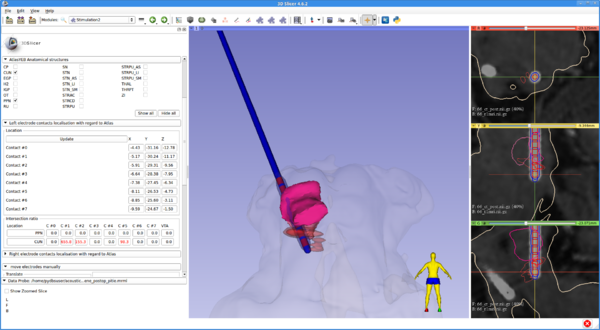
PyDBS is an automated image processing workflow for planning and postoperative assessment of deep brain stimulation interventions. It takes as input patient-specific data (i.e. patient images and patient clinical data) and generic models (i.e. an anatomical atlas, a model of the stereotactic frame and a model of the implanted electrodes) and provides as output a patient-specific model for planning and postoperative assessment of DBS surgery. This patient-specific model is composed of patient images, segmented anatomical structures (volumetric binary masks and triangular surface meshes) and geometrical transformations (registration matrices and deformation fields). All images, masks and meshes are mapped to a common reference space and fused in geometrical 3D scenes that can be readily visualized by the surgeon.
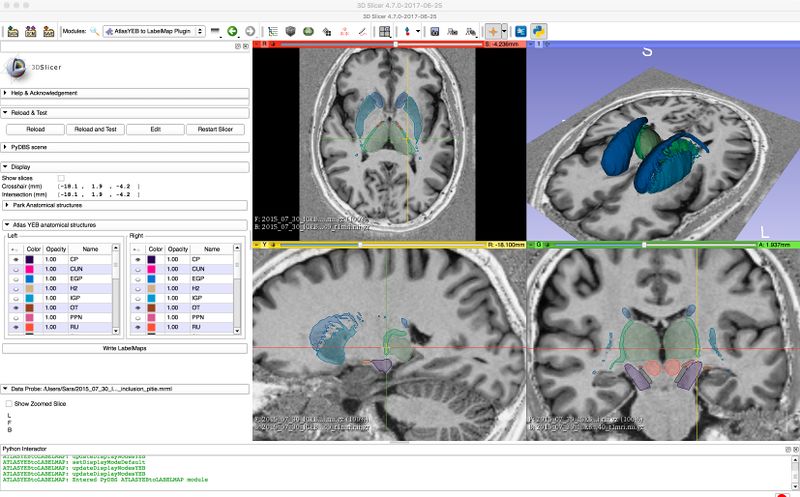
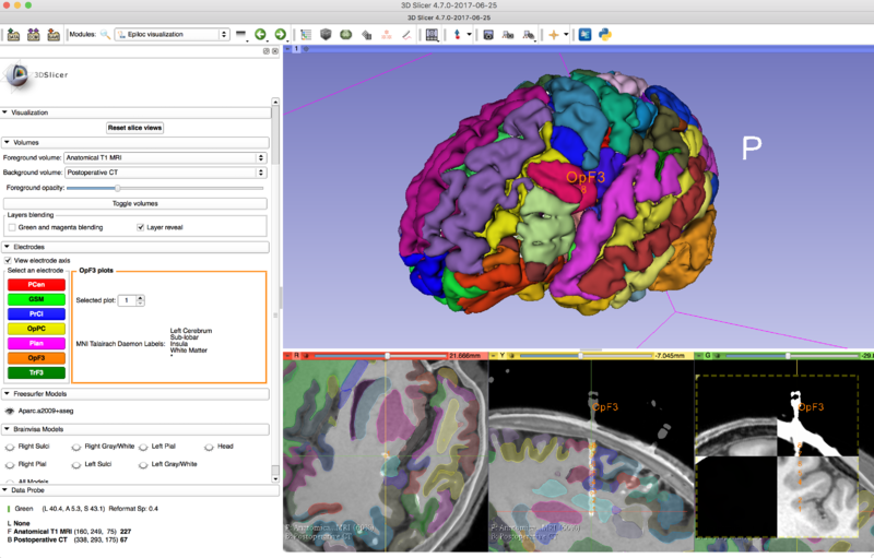
The software used for visualization and surgical planning is 3D Slicer. PyDBS includes several modules used for targeting and surgery assessment.
| Objective | Approach and Plan | Progress and Next Steps |
|---|---|---|
|
|
|


