Difference between revisions of "Projects:RegistrationTestbed"
Jfishbaugh (talk | contribs) |
m (Text replacement - "http://www.slicer.org/slicerWiki/index.php/" to "https://www.slicer.org/wiki/") |
||
| (13 intermediate revisions by one other user not shown) | |||
| Line 23: | Line 23: | ||
==Registration Testbed== | ==Registration Testbed== | ||
| − | [[ | + | Our proposed framework is similar in spirit to that of [http://www.batchmake.org Batchmake]. At the highest level of abstraction, the testbed is a common interface to command line registration applications. It allows for batch processing large experiments for single or multiple registration pipelines. We use the term pipeline to describe a collection of registration applications that are associated with each other. For example, the [http://www.doc.ic.ac.uk/~dr/software/ IRTK] pipeline consists of three applications: one for linear registration, one for non-linear registration, and finally an application to produce the output image. While there are three separate applications, they are used together for a single registration task. A pipeline in our testbed consists of one or more registration applications. |
| − | + | [[Image:Diagram1.png|thumb|center|300px|High level architecture.]] | |
At a high level, the testbed has three main components. First, the database represents a collection of definitions of known registration pipelines. This includes knowledge of required and optional input parameters and format of external parameter files used as input. The definitions allow for the implicit enforcement of any rules a given application might have. For example, an application might require a target and source image as the first two arguments, or optional parameters be specified with a "-" or "--". In other words, the database of known definitions helps distance a user from application specific details. This way, a user can more easily generate experiments with one or more registration pipelines without having to know how to use that specific application or consult various documentation. Any command line based registration application can be easily added to the database. | At a high level, the testbed has three main components. First, the database represents a collection of definitions of known registration pipelines. This includes knowledge of required and optional input parameters and format of external parameter files used as input. The definitions allow for the implicit enforcement of any rules a given application might have. For example, an application might require a target and source image as the first two arguments, or optional parameters be specified with a "-" or "--". In other words, the database of known definitions helps distance a user from application specific details. This way, a user can more easily generate experiments with one or more registration pipelines without having to know how to use that specific application or consult various documentation. Any command line based registration application can be easily added to the database. | ||
| Line 31: | Line 31: | ||
Experiments are stored in an XML format. An experiment file consists of necessary experiment information, such as paths to executables and parameter values. The main testbed application receives an experiment file as input, and using the database, formats the correct command line calls to conduct the experiment. An advanced user can interact directly with the XML to develop experiments. However, a user interface is available to help automate the process. | Experiments are stored in an XML format. An experiment file consists of necessary experiment information, such as paths to executables and parameter values. The main testbed application receives an experiment file as input, and using the database, formats the correct command line calls to conduct the experiment. An advanced user can interact directly with the XML to develop experiments. However, a user interface is available to help automate the process. | ||
| − | [[Image:Xmlexample.png|thumb|300px|A simple experiment file.]] | + | [[Image:Xmlexample.png|thumb|center|300px|A simple experiment file.]] |
| + | |||
| + | ==Experiments== | ||
| − | + | We now compare registration using the ITK based [https://www.slicer.org/wiki/Modules:RegisterImages-Documentation-3.4 RegisterImages] module found in Slicer and the [http://www.doc.ic.ac.uk/~dr/software/ Image Registration Toolkit] (IRTK). | |
| − | + | ===Intra-Subject Brain MRI: Axial T1 Tumor Growth Assessment=== | |
We begin with a tumor growth study. We wish to register the follow up scan to the baseline in order to measure the evolution of the tumor. We perform a rigid registration with six degrees of freedom, three for rotation and | We begin with a tumor growth study. We wish to register the follow up scan to the baseline in order to measure the evolution of the tumor. We perform a rigid registration with six degrees of freedom, three for rotation and | ||
| − | three for translation. Our choice of a rigid transformation is driven by our goal of change assessment, we do not want to introduce scaling during registration. Data taken from the [http://na-mic.org/Wiki/index.php/Projects:RegistrationDocumentation:UseCaseExample1b Registration Case Library]. | + | three for translation. Our choice of a rigid transformation is driven by our goal of change assessment, we do not want to introduce scaling or non-linear deformation during registration. Data taken from the [http://na-mic.org/Wiki/index.php/Projects:RegistrationDocumentation:UseCaseExample1b Registration Case Library]. |
| + | |||
| + | [[Image:All_both.png|thumb|center|700px|Left) First scan. Bottom) Follow up scan.]] | ||
This is an interesting experiment for two reasons. First, tumor change assessment is a common clinical task. As such, there is a need for registration applications to produce high quality results | This is an interesting experiment for two reasons. First, tumor change assessment is a common clinical task. As such, there is a need for registration applications to produce high quality results | ||
| Line 44: | Line 48: | ||
This often manifests in poor alignment for rigid registration, or in a large amount of deformation in the area of difference in a non-linear registration. | This often manifests in poor alignment for rigid registration, or in a large amount of deformation in the area of difference in a non-linear registration. | ||
| − | We further observe | + | We further observe different bias fields present in the baseline and follow up images. As a result, we choose a similarity metric that is not sensitive to intensity difference, namely mutual information. We also note that the data dimensions are 128x128x56 for the baseline and 128x128x65 for the follow up. The alignment before registration is |
| + | shown below. | ||
| + | |||
| + | [[Image:tumor_before.gif|thumb|center|340px| Alignment before registration. First scan in gray, follow up scan in red.]] | ||
| + | |||
| + | We next register the data using IRTK. The results are shown below. | ||
| + | |||
| + | [[Image:rview_tumor_reg.gif|thumb|center|340px| After registration using IRTK.]] | ||
| + | |||
| + | ===Atrial Fibrillation Study=== | ||
| − | + | Atrial fibrillation is an abnormal heart rhythm caused by disorganized electric impulses on the atrial wall. This causes the heart to beat in a quivering motion rather than an organized, efficient beat. An effective treatment is ablation surgery, where the abnormal tissue of the atrium is destroyed, typically performed by inserting a catheter and scarring areas of abnormal electric activity. The ablation procedure scars the tissue, rendering it ineffective in transmitting electric signals. The reduction in electric activity allows the heart to beat normally. | |
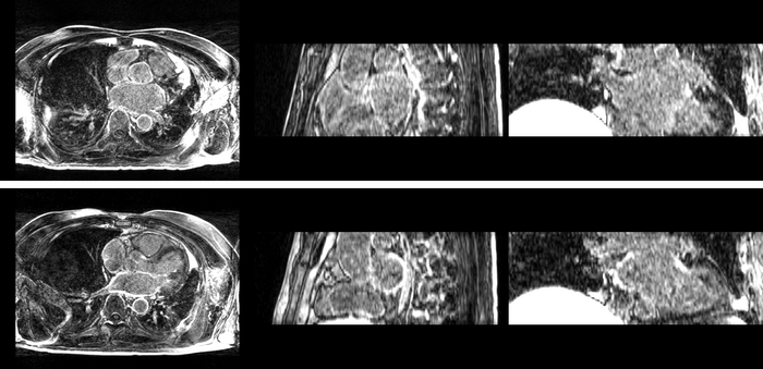
| − | [[Image: | + | [[Image:Prevspost.png|thumb|center|700px|Top) Pre-ablation. Bottom) Post-ablation.]] |
| − | + | The pre and post-ablation images are shown below. The data dimensions are 320x320x20 for the pre-ablation, and 288x288x20 for the post-ablation. We observe differences in shape present due to respiration and subject placement. Furthermore, the | |
| + | ablation procedure has scarred the tissue of the atrium, which can be seen in bright white in the post-ablation image. As a result, the shape of the atrium has likely changed. For these reasons, this registration task is particularly challenging. The alignment before registration is shown below. | ||
| − | + | [[Image:before.gif|thumb|center|700px| Alignment before registration. Pre-ablation in gray, post-ablation in red.]] | |
| − | [[Image: | + | [[Image:after.gif|thumb|center|700px|After registration using RegisterImages.]] |
| − | [[Image: | + | [[Image:Rview_after.gif|thumb|center|700px|After registration using IRTK.]] |
| − | [[Image: | + | [[Image:Registerimages_afib_roi_reg.gif|thumb|center|700px|After registration using RegisterImages.]] |
| − | [[Image: | + | [[Image:Rview_afib_roi_reg.gif|thumb|center|700px|After registration using IRTK.]] |
Latest revision as of 17:39, 10 July 2017
Home < Projects:RegistrationTestbedContents
Introduction
Image registration is a common step in medical imaging research pipelines. We must establish a common coordinate system across a set of images in order to perform meaningful analysis of the data.
A researcher interested in registering images has many options available. Dozens of algorithms for linear and non-linear registrations have been proposed and are well described in the literature. One possible option for the researcher is to implement the method they feel works best for their specific task. This works well for experts in registration, who are also proficient in software engineering. However, it is unclear how to systematically test registration for correctness. Testing with synthetic examples is possible, but we are ultimately interested in registration of real images. A useful method for testing/validation is comparison to existing registration applications.
Developing a custom registration tool is not a reasonable option for non-experts, for example a doctor performing a clinical study. A user in this case must choose an existing registration application. However, there are many registration packages available, many of which implement the same technique. It is difficult to know which algorithm is well suited for a given registration task, and furthermore, which implementation is best.
Most users fall into the category of non-experts in registration. They will likely use whatever registration package they are currently familiar with or seek the advice of a colleague. At this point, the user is exposed to another layer of difficulty -- parameter selection. It is difficult to select appropriate parameter values without knowledge of how image registration works. A frustrated user might conclude that the registration application they are using doesn't work, simply because they have not found suitable parameters for their data.
In response to the difficulties of image registration, we propose an environment where registration applications can be explored, tested, and compared. This registration testbed provides a common interface to several registration packages, allowing comparison experiments to be performed. A user can explore the impact of parameter settings by running experiments with varying parameter values. We also propose a collection of experiments representative of common registration task, such as multi-modal brain images, for benchmarking and comparison between registration packages.
Related Work
Slicer Registration Case Library
The Slicer Registration Case Library is an effort to establish a collection of registration examples with detailed documentation. The goal is to have a comprehensive collection of examples, so a users can refer to an example similar to their own registration task as a reference, including reasonable parameter settings.
Non-Rigid Image Registration Evaluation Project
The Non-Rigid Image Registration Evaluation Project (NIREP) is an evaluation project led by Gary Christensen at the University of Iowa. They are specifically interested in testing various deformable registration techniques, comparing them to existing registration applications such as SPM2, AIR, and ITK implementations. Furthermore, they propose the development of metrics for measuring the quality of the registration without knowledge of the true solution.
Registration Testbed
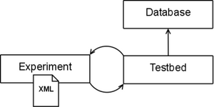
Our proposed framework is similar in spirit to that of Batchmake. At the highest level of abstraction, the testbed is a common interface to command line registration applications. It allows for batch processing large experiments for single or multiple registration pipelines. We use the term pipeline to describe a collection of registration applications that are associated with each other. For example, the IRTK pipeline consists of three applications: one for linear registration, one for non-linear registration, and finally an application to produce the output image. While there are three separate applications, they are used together for a single registration task. A pipeline in our testbed consists of one or more registration applications.
At a high level, the testbed has three main components. First, the database represents a collection of definitions of known registration pipelines. This includes knowledge of required and optional input parameters and format of external parameter files used as input. The definitions allow for the implicit enforcement of any rules a given application might have. For example, an application might require a target and source image as the first two arguments, or optional parameters be specified with a "-" or "--". In other words, the database of known definitions helps distance a user from application specific details. This way, a user can more easily generate experiments with one or more registration pipelines without having to know how to use that specific application or consult various documentation. Any command line based registration application can be easily added to the database.
Experiments are stored in an XML format. An experiment file consists of necessary experiment information, such as paths to executables and parameter values. The main testbed application receives an experiment file as input, and using the database, formats the correct command line calls to conduct the experiment. An advanced user can interact directly with the XML to develop experiments. However, a user interface is available to help automate the process.
Experiments
We now compare registration using the ITK based RegisterImages module found in Slicer and the Image Registration Toolkit (IRTK).
Intra-Subject Brain MRI: Axial T1 Tumor Growth Assessment
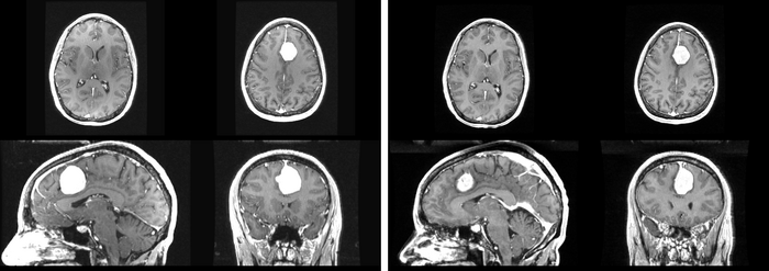
We begin with a tumor growth study. We wish to register the follow up scan to the baseline in order to measure the evolution of the tumor. We perform a rigid registration with six degrees of freedom, three for rotation and three for translation. Our choice of a rigid transformation is driven by our goal of change assessment, we do not want to introduce scaling or non-linear deformation during registration. Data taken from the Registration Case Library.
This is an interesting experiment for two reasons. First, tumor change assessment is a common clinical task. As such, there is a need for registration applications to produce high quality results for this type of data. Secondly, the presence of pathology makes registration challenging. If the difference between structures in the two images is considerable, registration algorithms will often produce results of poor quality. This often manifests in poor alignment for rigid registration, or in a large amount of deformation in the area of difference in a non-linear registration.
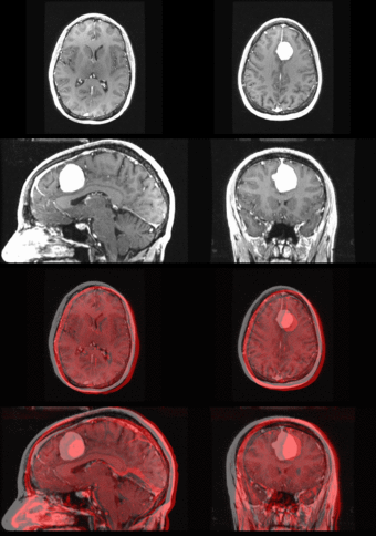
We further observe different bias fields present in the baseline and follow up images. As a result, we choose a similarity metric that is not sensitive to intensity difference, namely mutual information. We also note that the data dimensions are 128x128x56 for the baseline and 128x128x65 for the follow up. The alignment before registration is shown below.
We next register the data using IRTK. The results are shown below.
Atrial Fibrillation Study
Atrial fibrillation is an abnormal heart rhythm caused by disorganized electric impulses on the atrial wall. This causes the heart to beat in a quivering motion rather than an organized, efficient beat. An effective treatment is ablation surgery, where the abnormal tissue of the atrium is destroyed, typically performed by inserting a catheter and scarring areas of abnormal electric activity. The ablation procedure scars the tissue, rendering it ineffective in transmitting electric signals. The reduction in electric activity allows the heart to beat normally.
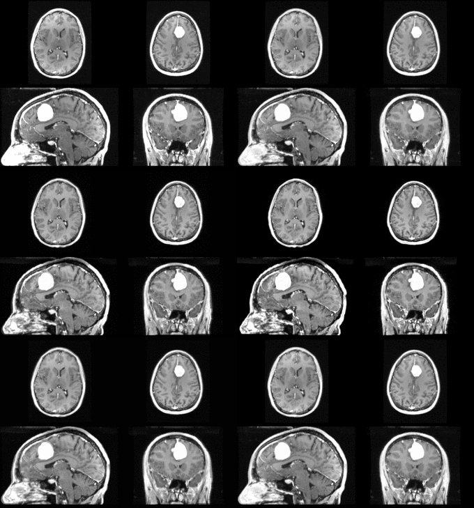
The pre and post-ablation images are shown below. The data dimensions are 320x320x20 for the pre-ablation, and 288x288x20 for the post-ablation. We observe differences in shape present due to respiration and subject placement. Furthermore, the ablation procedure has scarred the tissue of the atrium, which can be seen in bright white in the post-ablation image. As a result, the shape of the atrium has likely changed. For these reasons, this registration task is particularly challenging. The alignment before registration is shown below.
