Difference between revisions of "Mbirn: Machine Learning Tools"
m (Text replacement - "[http://www.na-mic.org/Wiki/images/" to "[https://na-mic.org/w/images/") |
|||
| Line 35: | Line 35: | ||
<br clear = "all" /> | <br clear = "all" /> | ||
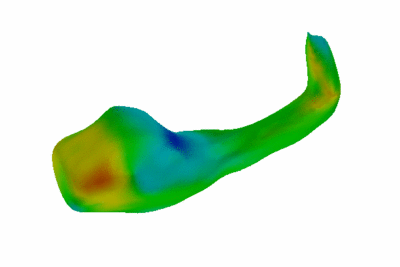
| − | <br /> In this study, we compared the shape of the hippocampus-amygdala complex of 15 schizophrenia patients and 15 normal controls. The classification accuracy achieved in a leave-one-out cross-valiadtion procedure was 76%. The figure above shows the direction (along the normal ot the surface) in which one would deform the normal hippocampus in the figure to make it look more like a schizophrenia hippocampus to the classifier function. Note that the units of deformation are not important, since the image illustrates the direction, not the margnitude of the deformation. The relative values of the local deformation show how different parts of the anatomical structure are deformed. This animation (zip'ed file) shows the deformation process: [ | + | <br /> In this study, we compared the shape of the hippocampus-amygdala complex of 15 schizophrenia patients and 15 normal controls. The classification accuracy achieved in a leave-one-out cross-valiadtion procedure was 76%. The figure above shows the direction (along the normal ot the surface) in which one would deform the normal hippocampus in the figure to make it look more like a schizophrenia hippocampus to the classifier function. Note that the units of deformation are not important, since the image illustrates the direction, not the margnitude of the deformation. The relative values of the local deformation show how different parts of the anatomical structure are deformed. This animation (zip'ed file) shows the deformation process: [https://na-mic.org/w/images/f/fe/HippocampalShapeDifferences.zip Click to view]. |
'''Publications''' | '''Publications''' | ||
Latest revision as of 18:27, 10 July 2017
Home < Mbirn: Machine Learning Tools
- Machine Learning Tool Goals
- Machine Learning Topics (P. Golland, T. Jaakkola)
- Automatic Hypothesis Generation
- Integration of Existing Tools into BIRN Portal (with Grethe, BIRN-CC)
Discriminative Shape Analysis
Our goal is to develop computational approaches to comparing populations based on the shape of anatomical structures. We use the discriminative framework to characterize the differences in shape by training a classifier function and studying its sensitivity to small perturbations in the input data. An additional benefit of employing the classification approach is that the resulting classifier function can be used to label new examples into one of the two populations, e.g., for early detection in population screening or prediction in longitudinal studies. We estimate the expected accuracy of the classifier in a jackknife procedure. We have also adapted a non-parametric permutation test to the classification setting to estimate the statistical significance of the detected differences and the observed classification accuracy.
Within mBIRN, we aim to provide the tools for the discriminative analysis and to integrate them within the computation infrastructure of BIRN portal. The classification tools can be applied to any set of features that represent distinct populations; they are not restricted to the shape descriptors.
Example: Study of Hippocampal Shape in Schizophrenia.
In this study, we compared the shape of the hippocampus-amygdala complex of 15 schizophrenia patients and 15 normal controls. The classification accuracy achieved in a leave-one-out cross-valiadtion procedure was 76%. The figure above shows the direction (along the normal ot the surface) in which one would deform the normal hippocampus in the figure to make it look more like a schizophrenia hippocampus to the classifier function. Note that the units of deformation are not important, since the image illustrates the direction, not the margnitude of the deformation. The relative values of the local deformation show how different parts of the anatomical structure are deformed. This animation (zip'ed file) shows the deformation process: Click to view.
Publications
B.T. T. Yeo, W. Ou, P. Golland. Invertible Filter Banks on the 2-Sphere. To Appear in Proceedings of Internation Conference in Image Processing, Atlanta, GA, October 2006. (pdf)
B.T. T. Yeo, W. Ou, P. Golland. Invertible Filter Banks on the 2-Sphere. Technical Report, February 2006. (pdf)
P. Golland, W.E.L. Grimson, M.E. Shenton, R. Kikinis. Detection and Analysis of Statistical Differences in Anatomical Shape. Medical Image Analysis, 9(1):69-86, 2005. (pdf)
P. Golland and B. Fischl. Permutation Tests for Classification: Towards Statistical Significance in Image-Based Studies. In Proc. IPMI'2003: The 18th International Conference on Information Processing and Medical Imaging, LNCS 2732:330-341, 2003.
P. Golland, B. Fischl, M. Spiridon, N. Kanwisher, R.L. Buckner, M.E. Shenton, R. Kikinis, A. Dale, W.E.L. Grimson. Discriminative Analysis for Image-Based Studies. In Proc. MICCAI'2002: Fifth International Conference on Medical Image Computing And Computer Assisted Intervention. LNCS 2488:508-515, 2002.
UPDATES
August 25, 2006 (P. Golland)
We are continuing development of explicit representations for shape and distribution of cortical folding. This is a joint effort with Bruce Fischl and is making extensive use of freesurfer tools. Our paper on performing feature detection on surfaces of spherical topology was accepted for publication at the International Conference on Image Processing; we will present the work in October. The paper extends a widely used paradigm of linear filtering to functions of a sphere and proves conditions for invertibility of filter banks when applied to such functions. The filters can be used to detect salient features of the folding and constructing population statistics of the features. Meanwhile, the work is underway to apply the methodology to cortical surfaces.
March 27, 2006 (P. Golland)
- Shape analysis scripts are available: Click here to download. The help files should be self-explanatory, and if they are not, please let us know. The scripts implement the algorithims described in the journal paper above.
- Work in progress is to make the scripts available to the mbirn community through the web portal.
November 18, 2005 (P. Golland)
- Identified a student to work on classification and visualization scripts. Our goal is to release these for use by all interested groups in mBIRN this winter.
September 20, 2005 (P. Golland, S. Pieper)
- In discussion with Steve Pieper, we identified the depression data from Duke as a potential second test bed. Got the recent paper on the data, working on getting access to the data set.
August 6, 2005 (T. Jaakkola)
- Research goals: capturing regularities in the patterns of variation of relevant subcortical regions by means of graphical models, and incorporating prior medical knowledge for the purpose of guiding the approaches to produce stronger medically relevant hypotheses. In other words, we proposed to adapt and apply graphical models to capture typical variations and expand the models to include patient records and other co-varying data sources.
- Research progress: Our initial focus has been on learning models from medical records or images translated into a discrete set of variable values. We have specifically focused on three key issues in this context: 1) appropriate use of predominantly incomplete records, 2) efficient calculations with models involving a large number of variables, and 3) incorporation of various alternative hypotheses about how variables may be related to each other. The work so far consists of primarily redesigning and rewriting software packages. Further progress is contingent on finding a graduate student.
July 30, 2005 (P. Golland)
- Statistical tests added to the scripts.
- Remaining tasks before the scripts can be released to other sites:
- add help for programmers (currently, the help files are targeted towards users);
- update the user manual based on the feedback from the initial testing;
- make the non-linear option available through the scripts.
- Need to identify another site to test the scripts. BWH might be a good candidate. Discuss with Jorge and Steve Pieper.
May 17, 2005 (P. Golland)
- The first version of scripts was created and tested at MGH on the hippocampal data set, using different representations of shape.
- The current set of scripts allow the input data to be in one of the two formats: raw binary (float) and ascii. One file per subject must be created.
February 2, 2005 (P. Golland)
- Together with Bruce Fischl, we identified a path towards integration of the classification tools into BIRN.
- We will create a set of shell scripts that will read a small number of environment variables to identify the subjects in the study and the data files. The scripts will then run all the components of the discriminative analysis:
- train a classifier function;
- create the visualization of the detected differences;
- cross-validate and run permutation testing.
- We will test the scripts locally, at MGH, on the hippocampal data from the Freesurfer segmentation.
- We will refine the scripts and release them to other sites.


