Difference between revisions of "Slicer:Workshops:User Training 101 SPujol"
From NAMIC Wiki
m (Text replacement - "http://www.slicer.org/slicerWiki/index.php/" to "https://www.slicer.org/wiki/") |
|||
| (6 intermediate revisions by 2 users not shown) | |||
| Line 1: | Line 1: | ||
| − | <div class="thumb tright"><div style="width: | + | <div class="thumb tright"><div style="width: 90px">[[Image:Nac.png|[[Image:Nac.png|Link to the NAC website]]]]<div |
| + | class="thumbcaption">[http://www.spl.harvard.edu/nac Link to the NAC website]</div></div></div> | ||
= Introduction = | = Introduction = | ||
| − | This is a training compendium for neuroimage analysis using | + | This is a training compendium for neuroimage analysis using 3DSlicer. |
| − | + | ||
| + | This series of courses will teach you how to | ||
* load and view data | * load and view data | ||
* segment data and create 3D models | * segment data and create 3D models | ||
* perform Diffusion Tensor Imaging and fMRI analysis | * perform Diffusion Tensor Imaging and fMRI analysis | ||
| − | For an overview, dowload this [[Media:NA-MIC-05-Slicer-Overview.ppt| slideshow]] | + | For an overview of Slicer, dowload this [[Media:NA-MIC-05-Slicer-Overview.ppt| slideshow]] |
| + | |||
| + | = Software = | ||
| + | <div class="floatleft"><span>[[Image:3DSlicer.png|80px|[[Image:3DSlicer.png| 3D Slicer Logo]]]]</span></div> | ||
| + | |||
| + | The current version of Slicer that is fully supported is Slicer 2.6. | ||
| + | |||
| + | Please follow the [https://www.slicer.org/wiki/Slicer:Slicer2.6_Getting_Started Slicer 2.6 Getting Started] instructions to install the version of the Slicer program appropriate to your platform. | ||
| + | |||
| + | |||
| + | |||
= Training Compendium = | = Training Compendium = | ||
| + | |||
For course related questions, please send an e-mail to Sonia Pujol, Ph.D. (spujol at bwh.harvard.edu). | For course related questions, please send an e-mail to Sonia Pujol, Ph.D. (spujol at bwh.harvard.edu). | ||
| − | |||
| − | |||
| − | |||
| − | |||
| − | |||
| − | |||
| − | |||
| − | |||
| − | |||
| − | |||
| − | |||
| − | |||
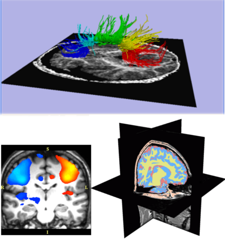
| − | = | + | [[Image:101.png|thumb|right|450px|Excerpts from the Slicer 101 courses: Diffusion Tensor Imaging fiber tractography (top); fMRI activation map (lower-left); automatic brain segmentation (lower-right).]] |
| − | + | ||
| − | + | {| border="2" | |
| − | + | |- bgcolor="white" | |
| − | + | | '''''' | |
| − | + | | '''Course''' | |
| − | + | | '''Dataset''' | |
| − | + | |- bgcolor="white" | |
| − | + | ||[[Image:Training1 LoadingViewing.PNG|50px]] | |
| − | + | | | |
| − | + | [[Media:SlicerTraining1LoadingAndViewingData.ppt| Data Loading and Visualization ]] | |
| − | + | | | |
| + | [[Media:Tutorial-with-dicom.zip|Tutorial_with_dicom.zip]] | ||
| + | |- bgcolor="white" | ||
| + | | [[Image:Training7Save.PNG|50px]] | ||
| + | | | ||
| + | [[Media:SlicerTraining7SavingData.ppt| Data Saving ]] | ||
| + | | | ||
| + | [[Media:Tutorial-with-dicom.zip|Tutorial-with-dicom.zip]] | ||
| + | |- bgcolor="white" | ||
| + | |[[Image:Training2Segmentation.PNG|50px]] | ||
| + | | | ||
| + | [[Media:Slicer_Segmentation_Tutorial.ppt| Manual Segmentation ]] | ||
| + | | | ||
| + | [[Media:Tutorial-with-dicom.zip|Tutorial_with_dicom.zip]] | ||
| + | |- bgcolor="white" | ||
| + | |[[Image:Training3LevelSets.PNG|50px]] | ||
| + | | | ||
| + | [[Media:03-LevelSet.ppt|Level-Set Segmentation ]] | ||
| + | | | ||
| + | [[Media:Tutorial-with-dicom.zip|Tutorial_with_dicom.zip]] | ||
| + | |- bgcolor="white" | ||
| + | | [[Image:Training10EM.png|50px]] | ||
| + | | | ||
| + | [[Media:SlicerAdvancedTraining_EMBrainAtlasClassifier_V1.0.ppt| Automatic Brain Segmentation ]] | ||
| + | | | ||
| + | [[Media:BrainAtlasClassifier.zip|BrainAtlasClassifier.zip ]] | ||
| + | |- bgcolor="white" | ||
| + | | [[Image:Training11Registration.PNG|55px]] | ||
| + | | | ||
| + | [[Media:SlicerAdvancedTraining11_Registration.ppt| Registration]] | ||
| + | | | ||
| + | [[Media:RegistrationSample.zip| RegistrationSample.zip]] | ||
| + | |- bgcolor="white" | ||
| + | |[[Image:Training4DTI.PNG|50px]] | ||
| + | | | ||
| + | [[Media:Slicer_DTMRI_Training4.ppt| Diffusion Tensor Imaging Analysis ]] | ||
| + | | | ||
| + | [[Media:SlicerSampleDTI.zip| SlicerSampleDTI.zip]] [[Media:Dwi-dicom.zip|Dwi-dicom.zip]] | ||
| + | |- bgcolor="white" | ||
| + | | [[Image:Training8Nrrd.PNG|50px]] | ||
| + | | | ||
| + | [[Media:SlicerTraining8-NrrdFileFormat.ppt| Nrrd File Format]] | ||
| + | | | ||
| + | [[Media:Tensor_data.zip|Tensor_data.zip]] | ||
| + | |- bgcolor="white" | ||
| + | | [[Image:Training9DicomToNrrd.PNG|50px]] | ||
| + | | | ||
| + | [[Media:SlicerTraining9_DTI-FromDicomToNrrd.ppt| Nrrd to Dicom Conversion ]] | ||
| + | | | ||
| + | [[Media:Dwi-dicom.zip|Dwi-dicom.zip]] | ||
| + | |- bgcolor="white" | ||
| + | | [[Image:Training5fMRI.PNG|50px]] | ||
| + | | | ||
| + | [[Media:SlicerTraining5_fMRI.ppt| Functional Magnetic Resonace Imaging Analysis ]] | ||
| + | | | ||
| + | [[Media:FMRIData1_short.zip|FMRIData1_short.zip]] [[Media:FMRIData2_long.zip|FMRIData2_long.zip]] | ||
| + | |- bgcolor="white" | ||
| + | | [[Image:Training6FreeSurfer.PNG|50px]] | ||
| + | | | ||
| + | [[Media:SlicerTraining6_vtkFreeSurferReaders.ppt| FreeSurfer Reader ]] | ||
| + | | | ||
| + | [[Media:FreeSurferSubjects.zip|FreeSurferSubjects.zip ]] | ||
| + | |} | ||
| − | |||
| − | |||
= Slicer Additional Resources = | = Slicer Additional Resources = | ||
| Line 46: | Line 107: | ||
* [[BrainAtlas|SPL PNL Brain Atlas]] | * [[BrainAtlas|SPL PNL Brain Atlas]] | ||
* [[IGT-Tutorials|IGT Tutorial Materials]] | * [[IGT-Tutorials|IGT Tutorial Materials]] | ||
| − | * [[Media:Training_EMLocalSegment_v1.pdf | EMSegmenter]] for multi-channel data and white matter hyperintensities | + | * [[Media:Training_EMLocalSegment_v1.pdf | EMSegmenter]] for multi-channel [[Media:EMSegmentTutorial.zip|data]] and white matter hyperintensities |
* [[DataFusion|Data Fusion and Registration]] | * [[DataFusion|Data Fusion and Registration]] | ||
* [http://astromed.iic.harvard.edu/UsingSlicer Using 3DSlicer in Astronomy] | * [http://astromed.iic.harvard.edu/UsingSlicer Using 3DSlicer in Astronomy] | ||
| − | * [ | + | * [https://www.slicer.org/wiki/SlicerOnBIRN How to use Slicer with BIRN] |
* [[media:Abdomen.zip|CT based atlas of the abdomen]]. This data is organized in analogy to the brain atlas above. The zip archive contains a .txt file with the key for the label values and their anatomical meaning. | * [[media:Abdomen.zip|CT based atlas of the abdomen]]. This data is organized in analogy to the brain atlas above. The zip archive contains a .txt file with the key for the label values and their anatomical meaning. | ||
| − | = Feedback | + | = Feedback = |
| − | * [ | + | * [http://www.slicer.org/pages/Slicer_Community Feedback about Slicer] |
* [http://www.na-mic.org/Bug/ Bug reports] | * [http://www.na-mic.org/Bug/ Bug reports] | ||
Latest revision as of 18:07, 10 July 2017
Home < Slicer:Workshops:User Training 101 SPujolContents
Introduction
This is a training compendium for neuroimage analysis using 3DSlicer.
This series of courses will teach you how to
- load and view data
- segment data and create 3D models
- perform Diffusion Tensor Imaging and fMRI analysis
For an overview of Slicer, dowload this slideshow
Software
The current version of Slicer that is fully supported is Slicer 2.6.
Please follow the Slicer 2.6 Getting Started instructions to install the version of the Slicer program appropriate to your platform.
Training Compendium
For course related questions, please send an e-mail to Sonia Pujol, Ph.D. (spujol at bwh.harvard.edu).
| ' | Course | Dataset |

|
||

|
||
Slicer Additional Resources
- SPL PNL Brain Atlas
- IGT Tutorial Materials
- EMSegmenter for multi-channel data and white matter hyperintensities
- Data Fusion and Registration
- Using 3DSlicer in Astronomy
- How to use Slicer with BIRN
- CT based atlas of the abdomen. This data is organized in analogy to the brain atlas above. The zip archive contains a .txt file with the key for the label values and their anatomical meaning.
Feedback
Acknowledgements
- Slicer is being developed by a community of contributors. For more information see the Slicer website.
Back to Training:Main


