Difference between revisions of "ProjectWeek200706:vtkITKWrapperForRuleBasedSegmentation"
m (Projects/Structural/2007 Project Week vtkITK Wrapper for Rule Based Segmentation moved to ProjectWeek200706:vtkITKWrapperForRuleBasedSegmentation) |
|||
| (18 intermediate revisions by 3 users not shown) | |||
| Line 1: | Line 1: | ||
{| | {| | ||
|[[Image:ProjectWeek-2007.png|thumb|320px|Return to [[2007_Programming/Project_Week_MIT|Project Week Main Page]] ]] | |[[Image:ProjectWeek-2007.png|thumb|320px|Return to [[2007_Programming/Project_Week_MIT|Project Week Main Page]] ]] | ||
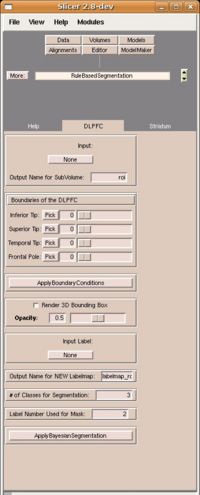
| − | |[[Image: | + | |[[Image:Rulebasedseg1.jpg|thumb|200px|The Slicer2 RuleBasedSegmentation module.]] |
| − | |||
|} | |} | ||
__NOTOC__ | __NOTOC__ | ||
===Key Investigators=== | ===Key Investigators=== | ||
| + | * Georgia Tech: Tauseef Rehman, John Melonakos | ||
* Kitware: Brad Davis | * Kitware: Brad Davis | ||
| − | * BWH: | + | * BWH: Nicole Aucoin, Marek Kubicki |
<div style="margin: 20px;"> | <div style="margin: 20px;"> | ||
<div style="width: 27%; float: left; padding-right: 3%;"> | <div style="width: 27%; float: left; padding-right: 3%;"> | ||
| + | |||
<h1>Objective</h1> | <h1>Objective</h1> | ||
| − | We are developing | + | We are developing rule-based segmentation techniques which speedup the process and improve the accuracy for delineating the DLPFC in brain MRI scans. Our objective is to develop Slicer modules to facilitate clinical use of these techniques. |
| − | |||
| + | A functional Slicer2 module has been developed and now needs to be tested and used by our Core 3 partners. In this project, we are supporting our Core 3 partners by making enhancements to improve the user friendliness of the module. | ||
</div> | </div> | ||
<div style="width: 27%; float: left; padding-right: 3%;"> | <div style="width: 27%; float: left; padding-right: 3%;"> | ||
| − | <h1> | + | <h1>Approach, Plan </h1> |
| − | + | Our approach for segmenting the DLPFC is described in the references below. The challenge is to make this software user-friendly to enable clinical use of the tool. Our plan for the week is to fix/add several features to improve the user experience. | |
| − | Our approach for | ||
</div> | </div> | ||
| Line 29: | Line 29: | ||
<h1>Progress</h1> | <h1>Progress</h1> | ||
| + | ====June 2007 Project Week==== | ||
| + | |||
| + | The Slicer2 RuleBasedSegmentation Module was troubleshooted to locate problems in the interface. The following problems were identified: | ||
| + | |||
| + | 1. The sliders to select the ROI bounds were not linked to the respective "Pick" buttons. | ||
| + | |||
| + | 2. There was no visual feedback to the user for completion of "ApplyBoundaryConditions" and "ApplyBayesianSegmentation" | ||
| + | commands. | ||
| + | |||
| + | 3. The 3D cube to initialize the ROI needed to be deselected after ROI selection. | ||
| + | |||
| + | 4. User could not select filter parameters such as number of classes, label number of the ROI mask, and the output filename. | ||
| + | |||
| + | 5. The filter output was not getting displayed correctly. | ||
| + | 6. The procedure could not be run multiple times. | ||
| − | |||
| − | |||
| − | ==== | + | All the above problems were fixed. The filter parameters (number of classes,label number of ROI mask) were exposed to the user through the GUI and set to common default values. The visual confirmation for the "ApplyBoundaryCondtions" was set to disabling the Bounding Cube and for the "ApplyBayesianSegmentation" the segmentation results were displayed correctly. The help tab and tooltip were updated to incorporate the UI changes. Multiple test runs were performed to confirm consistent behavior. |
| − | + | ||
| + | ====2005-2007==== | ||
| + | This code was developed between 2005-2007. First is was developed and tested in Matlab. Then the sub-volume creation rules were ported to Slicer2 while the Bayesian segmentation was ported to ITK (see the references below for more detail). Finally, in early 2007, a vtk wrapper of the ITK Bayesian code was developed, thus completing the Slicer2 RuleBasedSegmentation module. | ||
</div> | </div> | ||
| Line 44: | Line 59: | ||
| − | === | + | ===References=== |
| − | * | + | * Ramsey Al-Hakim, James Fallon, Delphine Nain, John Melonakos, and Allen Tannenbaum. A dorsolateral prefrontal cortex semi-automatic segmenter. In SPIE Medical Imaging, 2006. |
| − | * | + | * J. Melonakos, K. Krishnan, and A. Tannenbaum. An ITK Filter for Bayesian Segmentation: itkBayesianClassifierImageFilter. Insight Journal, 2006. |
| − | * | + | * J. Melonakos, R. Al-Hakim, J. Fallon, and A. Tannenbaum. Knowledge-Based Segmentation of Brain MRI Scans Using the Insight Toolkit. Insight Journal, 2005. |
| − | |||
Latest revision as of 14:52, 4 December 2007
Home < ProjectWeek200706:vtkITKWrapperForRuleBasedSegmentation Return to Project Week Main Page |
Key Investigators
- Georgia Tech: Tauseef Rehman, John Melonakos
- Kitware: Brad Davis
- BWH: Nicole Aucoin, Marek Kubicki
Objective
We are developing rule-based segmentation techniques which speedup the process and improve the accuracy for delineating the DLPFC in brain MRI scans. Our objective is to develop Slicer modules to facilitate clinical use of these techniques.
A functional Slicer2 module has been developed and now needs to be tested and used by our Core 3 partners. In this project, we are supporting our Core 3 partners by making enhancements to improve the user friendliness of the module.
Approach, Plan
Our approach for segmenting the DLPFC is described in the references below. The challenge is to make this software user-friendly to enable clinical use of the tool. Our plan for the week is to fix/add several features to improve the user experience.
Progress
June 2007 Project Week
The Slicer2 RuleBasedSegmentation Module was troubleshooted to locate problems in the interface. The following problems were identified:
1. The sliders to select the ROI bounds were not linked to the respective "Pick" buttons.
2. There was no visual feedback to the user for completion of "ApplyBoundaryConditions" and "ApplyBayesianSegmentation" commands.
3. The 3D cube to initialize the ROI needed to be deselected after ROI selection.
4. User could not select filter parameters such as number of classes, label number of the ROI mask, and the output filename.
5. The filter output was not getting displayed correctly.
6. The procedure could not be run multiple times.
All the above problems were fixed. The filter parameters (number of classes,label number of ROI mask) were exposed to the user through the GUI and set to common default values. The visual confirmation for the "ApplyBoundaryCondtions" was set to disabling the Bounding Cube and for the "ApplyBayesianSegmentation" the segmentation results were displayed correctly. The help tab and tooltip were updated to incorporate the UI changes. Multiple test runs were performed to confirm consistent behavior.
2005-2007
This code was developed between 2005-2007. First is was developed and tested in Matlab. Then the sub-volume creation rules were ported to Slicer2 while the Bayesian segmentation was ported to ITK (see the references below for more detail). Finally, in early 2007, a vtk wrapper of the ITK Bayesian code was developed, thus completing the Slicer2 RuleBasedSegmentation module.
References
- Ramsey Al-Hakim, James Fallon, Delphine Nain, John Melonakos, and Allen Tannenbaum. A dorsolateral prefrontal cortex semi-automatic segmenter. In SPIE Medical Imaging, 2006.
- J. Melonakos, K. Krishnan, and A. Tannenbaum. An ITK Filter for Bayesian Segmentation: itkBayesianClassifierImageFilter. Insight Journal, 2006.
- J. Melonakos, R. Al-Hakim, J. Fallon, and A. Tannenbaum. Knowledge-Based Segmentation of Brain MRI Scans Using the Insight Toolkit. Insight Journal, 2005.
