Difference between revisions of "Projects:CorpusFiberClustering"
| (3 intermediate revisions by the same user not shown) | |||
| Line 1: | Line 1: | ||
| + | Back to [[NA-MIC_Collaborations|NA-MIC_Collaborations]], [[Algorithm:MIT|MIT Algorithms]] | ||
| + | |||
= Corpus Fiber Clustering = | = Corpus Fiber Clustering = | ||
| − | + | Our objective is to measure differences in fractional anisotropy (FA), trace, mode, and volume of five subdivisions of the corpus callosum of schizophrenic patients vs. normal controls using an automatic clustering method and high resolution 3 Tesla data. | |
| − | |||
| − | |||
| − | |||
| − | |||
| − | |||
| − | + | = Description = | |
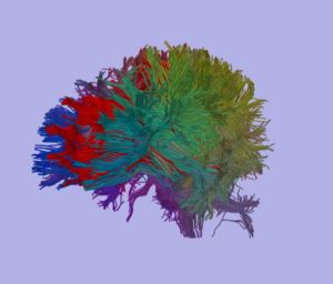
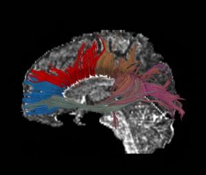
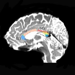
We are using a high resolution 3 Tesla scans, 1.7x1.7x1.7 mm, 51 diffusion directions, 8 baselines (data available to NAMIC investigators). So far we scanned 24 subjects, 12 chronic schizophrenics and 12 matched control subjects. Fiber tractography is performed on the whole brain, and then an automatic clustering method (O’Donnell et al., 2006), is applied. Midsagittal slice is then painted according to anatomical clusters, which results in 5, reliable clusters. Mean FA, mode, trace as well as volume of each clusters are obtained for each subject, and compared between groups. | We are using a high resolution 3 Tesla scans, 1.7x1.7x1.7 mm, 51 diffusion directions, 8 baselines (data available to NAMIC investigators). So far we scanned 24 subjects, 12 chronic schizophrenics and 12 matched control subjects. Fiber tractography is performed on the whole brain, and then an automatic clustering method (O’Donnell et al., 2006), is applied. Midsagittal slice is then painted according to anatomical clusters, which results in 5, reliable clusters. Mean FA, mode, trace as well as volume of each clusters are obtained for each subject, and compared between groups. | ||
| Line 16: | Line 13: | ||
[[image:clustering_brain.jpg|left|300px]] [[image:clustering_corpus.jpg|300px]] | [[image:clustering_brain.jpg|left|300px]] [[image:clustering_corpus.jpg|300px]] | ||
| + | [[image:clustering_corpus2.jpg|300px]] | ||
| − | + | = Publications = | |
| + | ''In Press'' | ||
| − | + | [http://www.na-mic.org/Special:Publications?text=Projects%3ACorpusFiberClustering&submit=Search&words=all&title=checked&keywords=checked&authors=checked&abstract=checked&sponsors=checked&searchbytag=checked| NA-MIC Publication Database] | |
| − | |||
| − | + | = Key Investigators = | |
| − | Harvard PNL: Marek Kubicki, Kate Smith, Martha Shenton | + | * Harvard PNL: Marek Kubicki, Kate Smith, Martha Shenton |
| − | MIT: Lauren O'Donnell, Carl-Fredrik Westin | + | * MIT: Lauren O'Donnell, Carl-Fredrik Westin |
Latest revision as of 18:46, 27 November 2007
Home < Projects:CorpusFiberClusteringBack to NA-MIC_Collaborations, MIT Algorithms
Corpus Fiber Clustering
Our objective is to measure differences in fractional anisotropy (FA), trace, mode, and volume of five subdivisions of the corpus callosum of schizophrenic patients vs. normal controls using an automatic clustering method and high resolution 3 Tesla data.
Description
We are using a high resolution 3 Tesla scans, 1.7x1.7x1.7 mm, 51 diffusion directions, 8 baselines (data available to NAMIC investigators). So far we scanned 24 subjects, 12 chronic schizophrenics and 12 matched control subjects. Fiber tractography is performed on the whole brain, and then an automatic clustering method (O’Donnell et al., 2006), is applied. Midsagittal slice is then painted according to anatomical clusters, which results in 5, reliable clusters. Mean FA, mode, trace as well as volume of each clusters are obtained for each subject, and compared between groups.
Preliminary results show a reduced FA in the second cluster (CC2) of the corpus callosum in schizophrenics and a reduced volume in the third cluster (CC3) of schizophrenics, when compared with control subjects.
Publications
In Press
Key Investigators
- Harvard PNL: Marek Kubicki, Kate Smith, Martha Shenton
- MIT: Lauren O'Donnell, Carl-Fredrik Westin


