Difference between revisions of "Downloads"
m (Text replacement - "http://www.slicer.org/slicerWiki/index.php/" to "https://www.slicer.org/wiki/") |
|||
| (79 intermediate revisions by 11 users not shown) | |||
| Line 1: | Line 1: | ||
| + | __TOC__ | ||
| + | |||
The following is a collection of electronic resources provided by NA-MIC. This includes software, data, tutorials, presentations, and additional documentation. | The following is a collection of electronic resources provided by NA-MIC. This includes software, data, tutorials, presentations, and additional documentation. | ||
==Software== | ==Software== | ||
| − | + | {|class=wikitable | |
| − | + | |[[Image:Slicer3_logo-i.jpg]] | |
| + | |[http://download.slicer.org Download Slicer]<br> | ||
| + | A general purpose biomedical computing application with extensive built-in visualization and analysis capabilities, accessible through an easy to use graphical interface. | ||
| + | |- | ||
| + | |[[Image:NAMIC-Kit-Overview-i.png]] | ||
| + | |[[NA-MIC-Kit | Download the NA-MIC Kit, including Slicer]]<br> | ||
| + | The NA-MIC Kit is a free open source software platform. The NA-MIC Kit is distributed under a BSD-style license without commercial restrictions or "give-back" requirements and is intended for research, but there are no restrictions on other uses. It consists of the 3D Slicer application software, a number of tools and toolkits such as VTK and ITK, and a software engineering methodology that enables multiplatform implementations. | ||
| + | |} | ||
==Data== | ==Data== | ||
| − | + | {|class=wikitable | |
| − | + | ||
| − | + | |[[Image:Schiz-thumb.png]] | |
| − | + | |[http://insight-journal.org/midas/collection/view/190 Brain: Multi-modality (sMRI, DTI, fMRI) from Schizophrenia Study]<br> | |
| − | + | There are 20 cases: ten are Normal Controls and ten are Schizophrenic. Each case includes a weighted T1 scan, a weighted T2 scan, an fMRI scan, a DTI volume, the DWI with 51 directions, and several masks and labelmaps. Available from Harvard. | |
| − | * [http://insight-journal.org/midas/ | + | |- |
| − | + | |[[Image:Child-i.png]] | |
| + | |[http://insight-journal.org/midas/community/view/24 Brain: 2-4 Year Old from Autism Study]<br> | ||
| + | Data for 2 autistic children and 2 normal controls (male, female) scanned at 2 years with follow up at 4 years from a 1.5T Siemens scanner. Files include structural data, tissue segmentation label map and subcortical structures segmentation. Available from UNC. | ||
| + | |- | ||
| + | |[[Image:Lupus-i.png]] | ||
| + | |[http://insight-journal.org/midas/collection/view/191 Brain: White Matter Lesions for Lupus Study]<br> | ||
| + | Data for 5 cases of Lupus White Matter Lesion patients. The data is co-registered. Each case contains: T1-weighted, T2-weighted, FLAIR, and masks for brain and lesions. Available from MIND. | ||
| + | |- | ||
| + | |[[Image:Prostate_thumb1.jpg|100px]] | ||
| + | |[http://insight-journal.org/midas/community/view/25 Prostate: 5 robot-assisted intervention cases for Prostate Cancer]<br> | ||
| + | MRI Prostate data. 5 datasets, with pre-operative and intra-operative scans (biopsy and seed placement procedure). Acquired at National Institute of Health (Principal Investigators: Camphausen, Kaushal and Pinto). | ||
| + | |- | ||
| + | |[[Image:Prostate_thumb2.jpg|100px]] | ||
| + | |[http://central.xnat.org/app/template/XDATScreen_report_xnat_projectData.vm/search_element/xnat:projectData/search_field/xnat:projectData.ID/search_value/NCIGT_PROSTATE Prostate: 10 cases]<br> | ||
| + | MRI Prostate data. 10 datasets, including a derived segmentation series with labelmaps. Available from Harvard. | ||
| + | |- | ||
| + | |[[Image:Trptutorial_thumb.jpg|100px]] | ||
| + | |[http://insight-journal.org/midas/collection/view/189 Prostate: Transrectal Tutorial Dataset]<br> | ||
| + | Transrectal Prostate Biopsy Tutorial Dataset. Walks the user through: Calibration (calibration image for the APT-MRI device), Segmentation (prostate MRI image and seeds for random walk segmentation algorithm), Targeting (target planning prostate MRI image), and Verification (needle insertion verification image). Available from Queens. | ||
| + | |- | ||
| + | |[[Image:Perkstation-i.jpg]] | ||
| + | |[http://insight-journal.org/midas/item/view/2454 Spine Phantom: PerkStation Tutorial Dataset]<br> | ||
| + | Perkstation Tutorial Dataset. Available from Queens. | ||
| + | |- | ||
| + | |[[Image:Vizhuman_thumb.png|100px]] | ||
| + | |[https://mri.radiology.uiowa.edu/visible_human_datasets.html Visible Human Datasets]<br> | ||
| + | Visible Human Datasets with some post-processing. Available from Iowa. | ||
| + | |- | ||
| + | |[[Image:SlicerRegistrationLibrary-i.png]] | ||
| + | |[[Projects:RegistrationDocumentation:UseCaseInventory | Registration Case Library Home Page]]<br> | ||
| + | New and growing (Oct. 2009 - Sept. 2011) list of image datasets for testing 3DSlicer registration methods & modules. Images range from brain to abdominal to musculoskeletal, modalities range from MRI, CT to PET. Data includes raw image data (NRRD), registration task description & discussion, results, parameter preset files and step-by step tutorials. Built for the clinician researcher to find a related image registration problem and thus provide a starting point for registration parameters and strategies. | ||
| + | *[http://www.insight-journal.org/midas/item/view/2332 Registration Library cases on the MIDAS database] | ||
| + | |- | ||
| + | |[[Image:CarmaData.png|100px]] | ||
| + | |[http://www.insight-journal.org/midas/collection/view/197 CARMA late-gadolinium MRI images and segmentations] <br> | ||
| + | Late-gadolinium enhancement data from the CARMA Center. Sixty anonymized sample datasets are currently available. They consist of pre-RF-ablation images and post-RF-ablation images along with manual segmentations of the left atrial walls, and MRA images as well. For most subjects, two post-RF-ablation images at 3 and 6 month intervals or 4 and 7 month intervals are available. Some subject datasets have post-RF-ablation images at different intervals and may not have MRA data. <br> | ||
| + | [http://www.insight-journal.org/midas/collection/view/197 CARMA Longitudinal Left Atrial Shape Data] <br> | ||
| + | Segmentations of the left atria of sixty anonymized sample datasets with the pulmonary veins and appendage removed. The segmentation data will be used for generating corresponding points and mappings between local and average left atrium shape using ShapeWorks, an open-source software with tools for preprocessing data, computing point-based shape models, and visualizing the results. | ||
| + | |} | ||
==Tutorials== | ==Tutorials== | ||
| + | '''Here is a sampling of the available tutorials for Biomedical Engineers and Clinical Research Users of the NA-MIC Kit (PDF and PPT downloads)''' | ||
| + | The full compendium is found [https://www.slicer.org/wiki/Slicer3.6:Training here] | ||
| + | {| class="wikitable" | ||
| + | |[[Image:Stochastic-i.png]] | ||
| + | |[[Media:Stochastic_June09_1.ppt|Stochastic Tractography to extract, visualize and quantify white matter connections from Diffusion Images in Schizophrenia Study]]<br> | ||
| + | The python stochastic tractography module contains the tools necessary to extract, visualize and quantify white matter connections from DTI images. It seeds nerve fiber bundles from regions of interest (ROIs) based on DWI images. Unlike streamline tractography, stochastic tractography uses a probabilistic framework to perform tractography. | ||
| + | |- | ||
| + | |[[Image:WhiteMatterLesions-i.png]] | ||
| + | |[[media:Slicer3Training_WhiteMatterLesions_v2.3.pdf|Classification of White Matter Lesions for Lupus]]<br> | ||
| + | This tutorial demonstrates an automated, multi-level method to segment white matter brain lesions in lupus. Following this tutorial, you’ll be able to load scans into Slicer3, and segment and measure the volume of white matter lesions on the provided data-set. | ||
| + | |- | ||
| + | |[[Image:TransRectal-i.png]] | ||
| + | |[[media:DBP2JohnsHopkinsTransRectalProstateBiopsy_TutorialPres2009June.pdf|Trans-rectal MR guided prostate biopsy]] and [[Media:PerkStationSlicerTutorial.pdf|PerkStationSlicerTutorial]]<br> | ||
| + | This tutorial will teach you how to perform MR-guided prostate biopsy using MR-compatible trans-rectal robot with SLICER. | ||
| + | |- | ||
| + | |[[Image:Arctic-i.png]] | ||
| + | |[[media:ARCTIC-Slicer3-Tutorial.ppt|ARCTIC: Automatic Regional Cortical ThICkness Analysis for Autism]]<br> | ||
| + | Following this tutorial, you will be able to perform an individual analysis of regional cortical thickness. You will learn how to load input volumes, run the end-to-end module ARCTIC to generate cortical thickness information and display output volumes. | ||
| + | |- | ||
| + | |[[Image:Confocal-i.png]] | ||
| + | |[[media:Microscopy-Confocal-TrainingTutorial-2009JUNE.pdf|Confocal Microscopy]]<br> | ||
| + | Guiding you step by step through the process of loading confocal microscopy data, working with that data, and creating a 3D model for visualization. | ||
| + | |- | ||
| + | |[[Image:Primate-i.jpg]] | ||
| + | |[[media:EMSegment_TrainingTutorial.pdf| Non-human Primates Segmentation Tutorial]]<br> | ||
| + | The objective of this tutorial is to demonstrate how to use EM Segmenter to segment non-human primate images. | ||
| + | |- | ||
| + | |[[Image:Hammer-i.png]] | ||
| + | |[[AHM 2010 Tutorial Contest - Hammer Registration | Hammer Registration for Brain MRI]]<br> | ||
| + | Presents HAMMER registration algorithm and introduces how to use HAMMER in Slicer3. | ||
| + | |- | ||
| + | |[[Image:CenterLine-i.png]] | ||
| + | |[[AHM 2010 Tutorial Contest - CoronaryArteriesCenterlinesVMTK | Centerline Extraction of Coronary Arteries using VMTK]]<br> | ||
| + | Guiding you step by step through the process of centerline extraction of Coronary Arteries in a cardiac blood-pool MRI using VMTK based Tools. | ||
| + | |- | ||
| + | |[[File:EMFiberClustering-i.png]] | ||
| + | |[[AHM 2010 Tutorial Contest - EM Fiber Clustering| EM Fiber Clustering]]<br> | ||
| + | This module clusters a set of input trajectories into a number of bundles, generates arc length parameterization by establishing the point correspondences and reports diffusion parameters along the bundles as well as the membership probability of each trajectory in each cluster. The module requires specification of seed trajectories (or initial centerlines) as representatives of the desired bundles. | ||
| + | |} | ||
| + | |||
| + | '''Additional Tutorials''' | ||
| − | * [ | + | *[https://www.slicer.org/wiki/Slicer3.6:Training Slicer Training] |
| − | |||
| − | |||
| − | |||
| − | |||
| − | |||
| − | |||
| − | |||
| − | |||
Latest revision as of 16:59, 10 July 2017
Home < DownloadsContents
The following is a collection of electronic resources provided by NA-MIC. This includes software, data, tutorials, presentations, and additional documentation.
Software
|
Download Slicer A general purpose biomedical computing application with extensive built-in visualization and analysis capabilities, accessible through an easy to use graphical interface. |
|
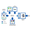
Download the NA-MIC Kit, including Slicer The NA-MIC Kit is a free open source software platform. The NA-MIC Kit is distributed under a BSD-style license without commercial restrictions or "give-back" requirements and is intended for research, but there are no restrictions on other uses. It consists of the 3D Slicer application software, a number of tools and toolkits such as VTK and ITK, and a software engineering methodology that enables multiplatform implementations. |
Data
|
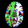
Brain: Multi-modality (sMRI, DTI, fMRI) from Schizophrenia Study There are 20 cases: ten are Normal Controls and ten are Schizophrenic. Each case includes a weighted T1 scan, a weighted T2 scan, an fMRI scan, a DTI volume, the DWI with 51 directions, and several masks and labelmaps. Available from Harvard. |
|
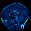
Brain: 2-4 Year Old from Autism Study Data for 2 autistic children and 2 normal controls (male, female) scanned at 2 years with follow up at 4 years from a 1.5T Siemens scanner. Files include structural data, tissue segmentation label map and subcortical structures segmentation. Available from UNC. |
|
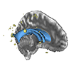
Brain: White Matter Lesions for Lupus Study Data for 5 cases of Lupus White Matter Lesion patients. The data is co-registered. Each case contains: T1-weighted, T2-weighted, FLAIR, and masks for brain and lesions. Available from MIND. |
|
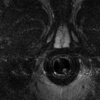
Prostate: 5 robot-assisted intervention cases for Prostate Cancer MRI Prostate data. 5 datasets, with pre-operative and intra-operative scans (biopsy and seed placement procedure). Acquired at National Institute of Health (Principal Investigators: Camphausen, Kaushal and Pinto). |
|
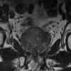
Prostate: 10 cases MRI Prostate data. 10 datasets, including a derived segmentation series with labelmaps. Available from Harvard. |

|
Prostate: Transrectal Tutorial Dataset Transrectal Prostate Biopsy Tutorial Dataset. Walks the user through: Calibration (calibration image for the APT-MRI device), Segmentation (prostate MRI image and seeds for random walk segmentation algorithm), Targeting (target planning prostate MRI image), and Verification (needle insertion verification image). Available from Queens. |
|
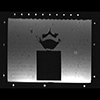
Spine Phantom: PerkStation Tutorial Dataset Perkstation Tutorial Dataset. Available from Queens. |
|
Visible Human Datasets Visible Human Datasets with some post-processing. Available from Iowa. |
|
Registration Case Library Home Page New and growing (Oct. 2009 - Sept. 2011) list of image datasets for testing 3DSlicer registration methods & modules. Images range from brain to abdominal to musculoskeletal, modalities range from MRI, CT to PET. Data includes raw image data (NRRD), registration task description & discussion, results, parameter preset files and step-by step tutorials. Built for the clinician researcher to find a related image registration problem and thus provide a starting point for registration parameters and strategies. |

|
CARMA late-gadolinium MRI images and segmentations Late-gadolinium enhancement data from the CARMA Center. Sixty anonymized sample datasets are currently available. They consist of pre-RF-ablation images and post-RF-ablation images along with manual segmentations of the left atrial walls, and MRA images as well. For most subjects, two post-RF-ablation images at 3 and 6 month intervals or 4 and 7 month intervals are available. Some subject datasets have post-RF-ablation images at different intervals and may not have MRA data. |
Tutorials
Here is a sampling of the available tutorials for Biomedical Engineers and Clinical Research Users of the NA-MIC Kit (PDF and PPT downloads) The full compendium is found here
|
Stochastic Tractography to extract, visualize and quantify white matter connections from Diffusion Images in Schizophrenia Study The python stochastic tractography module contains the tools necessary to extract, visualize and quantify white matter connections from DTI images. It seeds nerve fiber bundles from regions of interest (ROIs) based on DWI images. Unlike streamline tractography, stochastic tractography uses a probabilistic framework to perform tractography. |
|
Classification of White Matter Lesions for Lupus This tutorial demonstrates an automated, multi-level method to segment white matter brain lesions in lupus. Following this tutorial, you’ll be able to load scans into Slicer3, and segment and measure the volume of white matter lesions on the provided data-set. |
|
Trans-rectal MR guided prostate biopsy and PerkStationSlicerTutorial This tutorial will teach you how to perform MR-guided prostate biopsy using MR-compatible trans-rectal robot with SLICER. |
|
ARCTIC: Automatic Regional Cortical ThICkness Analysis for Autism Following this tutorial, you will be able to perform an individual analysis of regional cortical thickness. You will learn how to load input volumes, run the end-to-end module ARCTIC to generate cortical thickness information and display output volumes. |
|
Confocal Microscopy Guiding you step by step through the process of loading confocal microscopy data, working with that data, and creating a 3D model for visualization. |
|
Non-human Primates Segmentation Tutorial The objective of this tutorial is to demonstrate how to use EM Segmenter to segment non-human primate images. |

|
Hammer Registration for Brain MRI Presents HAMMER registration algorithm and introduces how to use HAMMER in Slicer3. |
|
Centerline Extraction of Coronary Arteries using VMTK Guiding you step by step through the process of centerline extraction of Coronary Arteries in a cardiac blood-pool MRI using VMTK based Tools. |
|
EM Fiber Clustering This module clusters a set of input trajectories into a number of bundles, generates arc length parameterization by establishing the point correspondences and reports diffusion parameters along the bundles as well as the membership probability of each trajectory in each cluster. The module requires specification of seed trajectories (or initial centerlines) as representatives of the desired bundles. |
Additional Tutorials