Difference between revisions of "Projects:AblationScarSegmentation"
| (20 intermediate revisions by the same user not shown) | |||
| Line 5: | Line 5: | ||
Atrial fibrillation is one of the most common heart conditions and can have very serious consequences such as stroke and heart failure. A technique called catheter radio-frequency (RF) ablation has recently emerged as a treatment. It involves burning the cardiac tissue that is responsible for the fibrillation. Even though this technique has been shown to work fairly well on atrial fibrillation patients, repeat procedures are often needed to fully correct the condition because surgeons lack the necessary tools to quickly evaluate the success of the procedure. | Atrial fibrillation is one of the most common heart conditions and can have very serious consequences such as stroke and heart failure. A technique called catheter radio-frequency (RF) ablation has recently emerged as a treatment. It involves burning the cardiac tissue that is responsible for the fibrillation. Even though this technique has been shown to work fairly well on atrial fibrillation patients, repeat procedures are often needed to fully correct the condition because surgeons lack the necessary tools to quickly evaluate the success of the procedure. | ||
| − | We propose a method to automatically segment the scar created by RF ablation in delayed enhancement MR images acquired after the procedure. This | + | We propose a method to automatically segment the scar created by RF ablation in delayed enhancement MR images acquired after the procedure. This will provide surgeons with a visualization showing the size, shape and location of the scar, which is information central to evaluating the outcome of the procedure. |
= Description = | = Description = | ||
| Line 11: | Line 11: | ||
We work with two types of images for each patient: MR angiography (MRA) images where the blood pool has a higher intensity than surrounding tissue and post-procedure delayed enhancement MR images (DE-MRI) where a contrast agent has been injected into the patient to enhance the ablation scar. Our approach is to first segment the left atrium in the MRA images using the label fusion algorithm described in [1]. We then transfer this segmentation to the DE-MRI image of the same patient by registering the two images. | We work with two types of images for each patient: MR angiography (MRA) images where the blood pool has a higher intensity than surrounding tissue and post-procedure delayed enhancement MR images (DE-MRI) where a contrast agent has been injected into the patient to enhance the ablation scar. Our approach is to first segment the left atrium in the MRA images using the label fusion algorithm described in [1]. We then transfer this segmentation to the DE-MRI image of the same patient by registering the two images. | ||
| − | Since the ablation scar we are trying to segment is known to be located on the left atrium myocardium, we use this spatial prior information to reduce the search space for the ablation scar to only a small vicinity of the left atrium surface. This avoids many false positives caused by the noise in the DE-MRI images. We | + | Since the ablation scar we are trying to segment is known to be located on the left atrium myocardium, we use this spatial prior information to reduce the search space for the ablation scar to only a small vicinity of the left atrium surface. This avoids many false positives caused by the noise in the DE-MRI images. We will be exploring different segmentation methods in this ongoing work. |
== Results == | == Results == | ||
| − | Here we present results we have obtained for one subject using our methods. In the following images, we show the left atrium segmentation in the MRA image as an outline in one slice as well as a 3D | + | Here we present results we have obtained for one subject using our methods. In the following images, we show the left atrium segmentation in the MRA image as an outline in one slice as well as a 3D model. |
{| | {| | ||
| Line 26: | Line 26: | ||
{| | {| | ||
| − | | [[Image:Mdepa_MRA_MDE_registration.png| | + | | [[Image:Mdepa_MRA_MDE_registration.png|320px]] |
| [[Image:Mdepa_MDE_la_segmentation.png|250px]] | | [[Image:Mdepa_MDE_la_segmentation.png|250px]] | ||
|} | |} | ||
| − | Finally, we show some preliminary results of our ablation scar segmentation obtained using intensity thresholding. | + | Finally, we show some preliminary results of our cardiac ablation scar segmentation obtained using this spatial prior knowledge and intensity thresholding. The figure on the right also shows an expert manual segmentation of the scar alongside our result. |
{| | {| | ||
| − | | [[Image:Mdepa_MDE_scar_segmentation.png| | + | | [[Image:Mdepa_MDE_scar_segmentation.png|300px]] |
| − | | [[Image:Mdepa_MDE_scar_seg_3D.png| | + | | [[Image:Mdepa_MDE_scar_seg_3D.png|350px]] |
|} | |} | ||
| − | |||
= Literature = | = Literature = | ||
| Line 44: | Line 43: | ||
= Key Investigators = | = Key Investigators = | ||
| − | *MIT: Michal Depa and Polina Golland | + | *MIT: [http://people.csail.mit.edu/mdepa/ Michal Depa] and Polina Golland |
*BWH: Ehud Schmidt and Ron Kikinis | *BWH: Ehud Schmidt and Ron Kikinis | ||
| + | |||
| + | = Publications = | ||
| + | |||
| + | [http://www.na-mic.org/publications/pages/display?search=Projects%3AAblationScarSegmentation&submit=Search&words=all&title=checked&keywords=checked&authors=checked&abstract=checked&sponsors=checked&searchbytag=checked| NA-MIC Publications Database on Left Atrium Segmentation] | ||
Latest revision as of 21:08, 30 March 2011
Home < Projects:AblationScarSegmentationBack to NA-MIC Collaborations, MIT Algorithms,
Cardiac ablation scar segmentation
Atrial fibrillation is one of the most common heart conditions and can have very serious consequences such as stroke and heart failure. A technique called catheter radio-frequency (RF) ablation has recently emerged as a treatment. It involves burning the cardiac tissue that is responsible for the fibrillation. Even though this technique has been shown to work fairly well on atrial fibrillation patients, repeat procedures are often needed to fully correct the condition because surgeons lack the necessary tools to quickly evaluate the success of the procedure.
We propose a method to automatically segment the scar created by RF ablation in delayed enhancement MR images acquired after the procedure. This will provide surgeons with a visualization showing the size, shape and location of the scar, which is information central to evaluating the outcome of the procedure.
Description
We work with two types of images for each patient: MR angiography (MRA) images where the blood pool has a higher intensity than surrounding tissue and post-procedure delayed enhancement MR images (DE-MRI) where a contrast agent has been injected into the patient to enhance the ablation scar. Our approach is to first segment the left atrium in the MRA images using the label fusion algorithm described in [1]. We then transfer this segmentation to the DE-MRI image of the same patient by registering the two images.
Since the ablation scar we are trying to segment is known to be located on the left atrium myocardium, we use this spatial prior information to reduce the search space for the ablation scar to only a small vicinity of the left atrium surface. This avoids many false positives caused by the noise in the DE-MRI images. We will be exploring different segmentation methods in this ongoing work.
Results
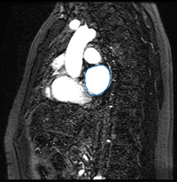
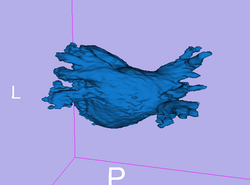
Here we present results we have obtained for one subject using our methods. In the following images, we show the left atrium segmentation in the MRA image as an outline in one slice as well as a 3D model.

|

|
We then align the MRA and DE-MRI images of the same patient, which allows us to transfer the left atrium segmentation using the resulting deformation field. This is shown in the following figures.

|

|
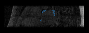
Finally, we show some preliminary results of our cardiac ablation scar segmentation obtained using this spatial prior knowledge and intensity thresholding. The figure on the right also shows an expert manual segmentation of the scar alongside our result.

|

|
Literature
[1] Nonparametric Mixture Models for Supervised Image Parcellation, M.R. Sabuncu, B.T.T. Yeo, K. Van Leemput, B. Fischl, and P. Golland. PMMIA Workshop at MICCAI 2009.
Key Investigators
- MIT: Michal Depa and Polina Golland
- BWH: Ehud Schmidt and Ron Kikinis