Difference between revisions of "2010 Summer Project Week Intraoperative Brain Shift Monitoring Using Shear Mode Transcranial Ultrasound"
From NAMIC Wiki
(Created page with '__NOTOC__ <gallery> Image:PW-MIT2010.png|Projects List Image:RSSMandible.png|Segmentation of the mandible using the RSS module. </gallery> …') |
|||
| (9 intermediate revisions by the same user not shown) | |||
| Line 2: | Line 2: | ||
<gallery> | <gallery> | ||
Image:PW-MIT2010.png|[[2010_Summer_Project_Week#Projects|Projects List]] | Image:PW-MIT2010.png|[[2010_Summer_Project_Week#Projects|Projects List]] | ||
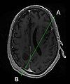
| − | Image: | + | Image:ITUM_NCIGT_PW.jpg|[[Fig. 1: Registration of Ultrasound Trajectory with MRI]] |
| + | Image:ITUM_SS.jpg|[[Fig. 2: ITUM Research Platform]] | ||
| + | Image:Slicer_SS.jpg|[[Fig. 3: Slicer with ITUM Data]] | ||
</gallery> | </gallery> | ||
==Key Investigators== | ==Key Investigators== | ||
| − | * | + | * BWH: Phillip Jason White, Alex Golby, Steve Pieper |
| − | |||
<div style="margin: 20px;"> | <div style="margin: 20px;"> | ||
| Line 13: | Line 14: | ||
<h3>Objective</h3> | <h3>Objective</h3> | ||
| − | + | Real-time ultrasound data is used to track the shifting of intracranial anatomy during neurosurgery. This is accomplished with a stationary ultrasound probe that is mounted away from the surgical site, and operates transcranially so as to be completely unobtrusive to surgical activities. Coordinating the use of this device with Slicer and BrainLab: | |
| − | * | + | * Registering the data obtained from it with pre-op images (MRI, CT, etc.) (Fig. 1) |
| + | * Using this data to modify/deform the pre-op images in order to maintain a more accurate representation of intraoperative anatomy. | ||
</div> | </div> | ||
| Line 21: | Line 23: | ||
<h3>Approach, Plan</h3> | <h3>Approach, Plan</h3> | ||
| − | |||
| − | |||
</div> | </div> | ||
| Line 29: | Line 29: | ||
<h3>Progress</h3> | <h3>Progress</h3> | ||
| − | + | Data transferred from ITUM Research Platform to Slicer via OpenIGTLink (Figs. 2-3) | |
| − | |||
</div> | </div> | ||
</div> | </div> | ||
| Line 38: | Line 37: | ||
==Delivery Mechanism== | ==Delivery Mechanism== | ||
| − | |||
==References== | ==References== | ||
</div> | </div> | ||
Latest revision as of 13:55, 25 June 2010
Home < 2010 Summer Project Week Intraoperative Brain Shift Monitoring Using Shear Mode Transcranial UltrasoundKey Investigators
- BWH: Phillip Jason White, Alex Golby, Steve Pieper
Objective
Real-time ultrasound data is used to track the shifting of intracranial anatomy during neurosurgery. This is accomplished with a stationary ultrasound probe that is mounted away from the surgical site, and operates transcranially so as to be completely unobtrusive to surgical activities. Coordinating the use of this device with Slicer and BrainLab:
- Registering the data obtained from it with pre-op images (MRI, CT, etc.) (Fig. 1)
- Using this data to modify/deform the pre-op images in order to maintain a more accurate representation of intraoperative anatomy.
Approach, Plan
Progress
Data transferred from ITUM Research Platform to Slicer via OpenIGTLink (Figs. 2-3)


