Difference between revisions of "DBP3:Utah:PV BFReg"
| (2 intermediate revisions by the same user not shown) | |||
| Line 3: | Line 3: | ||
== Pilot Study on BrainsFit Registration of LA Wall and Pulmonary Veins == | == Pilot Study on BrainsFit Registration of LA Wall and Pulmonary Veins == | ||
| − | + | Image registration is clinically useful for the alignment of medical images acquired at different time points (among a wide variety of other uses). For the CARMA Center, image registration of longitudinal DEMRI scans can help localize scar formation, the hallmark of ablation therapy success, or the lack thereof. We have experimented with using the BrainsFit registration framework from Slicer to register patient scans, allowing evaluating of lesion formation and lesion gaps around the PVs, a common ablation location. | |
=== Examples === | === Examples === | ||
| + | (click to enlarge) | ||
{| class="wikitable" | {| class="wikitable" | ||
| Line 13: | Line 14: | ||
! PV Misregistration | ! PV Misregistration | ||
|- | |- | ||
| − | | [[File:Good_Registration_CARMA.png|thumb]] | + | | [[File:Good_Registration_CARMA.png|thumb|center]] |
| − | | [[File:Septal_MisReg_CARMA.png|thumb]] | + | | [[File:Septal_MisReg_CARMA.png|thumb|center]] |
| − | | [[File:PV_MisReg_CARMA.png|thumb]] | + | | [[File:PV_MisReg_CARMA.png|thumb|center]] |
| + | |- | ||
| + | | [[File:Good_Checker_CARMA.png|thumb|center]] | ||
| + | | [[File:Septal_Checker_CARMA.png|thumb|center]] | ||
| + | | [[File:PV_Checker_CARMA.png|thumb|center]] | ||
|- | |- | ||
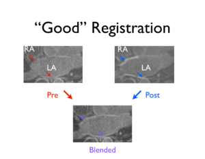
| An example of generally good registration of both the LA wall and PVs on a patient's pre- and 3 month post-ablation DEMRI scans (affine and rigid registration via BrainsFit module). | | An example of generally good registration of both the LA wall and PVs on a patient's pre- and 3 month post-ablation DEMRI scans (affine and rigid registration via BrainsFit module). | ||
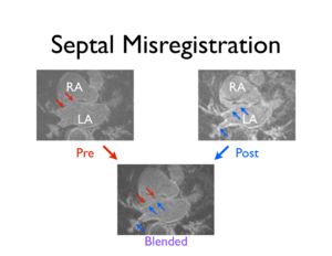
| An example of the pre- and post-ablation images from the same patient that were registered (affine and rigid) with the BrainsFit module. The registration was good except along the septal surface (arrows). | | An example of the pre- and post-ablation images from the same patient that were registered (affine and rigid) with the BrainsFit module. The registration was good except along the septal surface (arrows). | ||
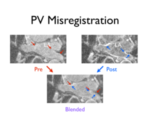
| − | | Affine and rigid registration (BrainsFit module) of the same patient's pre- and post-ablation scans improved the alignment of the PVs, but sufficient overlap of the corresponding PV structures was not achieved. | + | | Affine and rigid registration (BrainsFit module) of the same patient's pre- and post-ablation scans improved the alignment of the PVs, but sufficient overlap of the corresponding PV structures was not achieved (arrows). |
|- | |- | ||
|} | |} | ||
| Line 25: | Line 30: | ||
A short movie showing a linear blend between the septal misregistration images (fades from pre- to post-ablation scans). | A short movie showing a linear blend between the septal misregistration images (fades from pre- to post-ablation scans). | ||
| − | [[File:Septal_Reg_Movie_CARMA.mov | + | [[File:Septal_Reg_Movie_CARMA.mov]] |
| + | |||
| + | |||
| + | A second short clip showing a linear blend between the PV misregistration images above (again, from pre- to post-ablation scans). [[File:PV_Reg_Movie_CARMA.mov]] | ||
| + | |||
| + | === Initial Observations === | ||
| + | The BrainsFIt module seems to generally do a good job of aligning the posterior wall of the LA. The septal and anterior walls, which are more susceptible to motion during contraction, are more difficult to register correctly. The atrium wall is only a few pixels thick in the DEMRI images, so even small misalignments render comparisons between longitudinal scans inaccurate. The PVs have been difficult to register with just the affine and rigid routines provided by the BrainsFit module. However, the automatic image registration does get close to a complete registration of the scans, providing a jumping off point for more application-specific registration methods. | ||
| − | + | === Future Work === | |
| + | *Further experimentation with the affine and rigid transformations from the BrainsFit module | ||
| + | *Manually place fiducial markers in stable areas as a means of defining less fickle transformations at the LA surface | ||
| + | *Look at deformable transformations (such as B-spline in BrainsFit module) | ||
| + | *In conjunction with GA Tech, use the automatically generated LA segmentations to guide registration of the LA | ||
Latest revision as of 21:13, 9 November 2011
Home < DBP3:Utah:PV BFRegback to DBP3 home
Contents
The CARMA DBP: MRI-based study and treatment of atrial fibrillation
Pilot Study on BrainsFit Registration of LA Wall and Pulmonary Veins
Image registration is clinically useful for the alignment of medical images acquired at different time points (among a wide variety of other uses). For the CARMA Center, image registration of longitudinal DEMRI scans can help localize scar formation, the hallmark of ablation therapy success, or the lack thereof. We have experimented with using the BrainsFit registration framework from Slicer to register patient scans, allowing evaluating of lesion formation and lesion gaps around the PVs, a common ablation location.
Examples
(click to enlarge)
| Good LA Wall and PV Registration | Septal Misregistration | PV Misregistration |
|---|---|---|
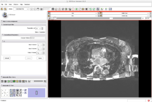
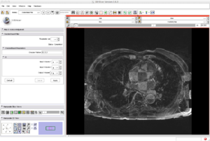
| An example of generally good registration of both the LA wall and PVs on a patient's pre- and 3 month post-ablation DEMRI scans (affine and rigid registration via BrainsFit module). | An example of the pre- and post-ablation images from the same patient that were registered (affine and rigid) with the BrainsFit module. The registration was good except along the septal surface (arrows). | Affine and rigid registration (BrainsFit module) of the same patient's pre- and post-ablation scans improved the alignment of the PVs, but sufficient overlap of the corresponding PV structures was not achieved (arrows). |
A short movie showing a linear blend between the septal misregistration images (fades from pre- to post-ablation scans).
A second short clip showing a linear blend between the PV misregistration images above (again, from pre- to post-ablation scans).
Initial Observations
The BrainsFIt module seems to generally do a good job of aligning the posterior wall of the LA. The septal and anterior walls, which are more susceptible to motion during contraction, are more difficult to register correctly. The atrium wall is only a few pixels thick in the DEMRI images, so even small misalignments render comparisons between longitudinal scans inaccurate. The PVs have been difficult to register with just the affine and rigid routines provided by the BrainsFit module. However, the automatic image registration does get close to a complete registration of the scans, providing a jumping off point for more application-specific registration methods.
Future Work
- Further experimentation with the affine and rigid transformations from the BrainsFit module
- Manually place fiducial markers in stable areas as a means of defining less fickle transformations at the LA surface
- Look at deformable transformations (such as B-spline in BrainsFit module)
- In conjunction with GA Tech, use the automatically generated LA segmentations to guide registration of the LA
