Difference between revisions of "2014 Project Week:CardiacCongenitalSegmentation"
From NAMIC Wiki
| (2 intermediate revisions by the same user not shown) | |||
| Line 3: | Line 3: | ||
Image:PW-SLC2014.png|[[2014_Winter_Project_Week#Projects|Projects List]] | Image:PW-SLC2014.png|[[2014_Winter_Project_Week#Projects|Projects List]] | ||
Image:Normal_vs_DORV_lrg.gif|Double outlet right ventricle | Image:Normal_vs_DORV_lrg.gif|Double outlet right ventricle | ||
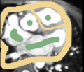
| + | Image:carreraScribbles.png|Example input to Carrera on axial slice | ||
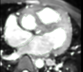
| + | Image:carreraOutput.png|Carrera output after manual interaction | ||
</gallery> | </gallery> | ||
| Line 30: | Line 32: | ||
<div style="width: 27%; float: left; padding-right: 3%;"> | <div style="width: 27%; float: left; padding-right: 3%;"> | ||
<h3>Progress</h3> | <h3>Progress</h3> | ||
| − | * | + | * Focused on blood pool segmentation in one test case. Tried: |
| + | ** CARMA tools: isolated connected / connected threshold operators | ||
| + | ** Editor level tracing effect | ||
| + | ** Editor Fast Marching effect | ||
| + | ** Editor Grow Cuts effect | ||
| + | ** Carrera interactive segmentation | ||
| + | ** Robust statistic active contour segmentation | ||
| + | * Best tool = Carrera interactive segmentation | ||
| + | ** Main difficulty = small chamber/vessel walls assigned as blood pool, but these can be fixed within Carrera somewhat easily | ||
| + | * Also tried affine registrations across my 5 subjects, using BRAINSFIT | ||
| + | ** Works very roughly, as expected | ||
| + | * Next steps: | ||
| + | ** Try Carrera on additional datasets for blood pool segmentation | ||
| + | ** Myocardium segmentation is still a challenge | ||
| + | * Thanks to: Josh Cates, Salma Bengali, Yi Gao, Ivan Kolesov for your help and suggestions! | ||
</div> | </div> | ||
</div> | </div> | ||
Latest revision as of 15:15, 10 January 2014
Home < 2014 Project Week:CardiacCongenitalSegmentationKey Investigators
Danielle Pace, MIT
Polina Golland, MIT
Project Description
Objective
- Develop a semi-automatic segmentation algorithm for cardiac MR images of patients with congenital heart defects.
- Goal is to build a surface model showing the endocardial and epicardial boundaries, for surgical planning.
- Challenges:
- Very large inter-subject variability due to heart defects
- Intensity inhomogeneities within myocardium and blood pool
- Similar intensity distributions within adjacent tissues (e.g. liver, chest muscle)
Approach, Plan
- Try existing open-source tools for segmentation and registration on the five datasets that we have so far.
- See where these methods fail, to focus our efforts for developing algorithms for segmenting hearts with congenital defects.
Progress
- Focused on blood pool segmentation in one test case. Tried:
- CARMA tools: isolated connected / connected threshold operators
- Editor level tracing effect
- Editor Fast Marching effect
- Editor Grow Cuts effect
- Carrera interactive segmentation
- Robust statistic active contour segmentation
- Best tool = Carrera interactive segmentation
- Main difficulty = small chamber/vessel walls assigned as blood pool, but these can be fixed within Carrera somewhat easily
- Also tried affine registrations across my 5 subjects, using BRAINSFIT
- Works very roughly, as expected
- Next steps:
- Try Carrera on additional datasets for blood pool segmentation
- Myocardium segmentation is still a challenge
- Thanks to: Josh Cates, Salma Bengali, Yi Gao, Ivan Kolesov for your help and suggestions!


