Difference between revisions of "2014 Summer Project Week:Ventriculostomy Guidance Transcranial Ultrasound"
From NAMIC Wiki
| (2 intermediate revisions by the same user not shown) | |||
| Line 2: | Line 2: | ||
<gallery> | <gallery> | ||
Image:PW-MIT2014.png|[[2014_Summer_Project_Week#Projects|Projects List]] | Image:PW-MIT2014.png|[[2014_Summer_Project_Week#Projects|Projects List]] | ||
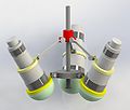
| − | + | Image:EVDGUI1.jpg|Transcranial ultrasound device for guiding the placement of an extra-ventricular drain | |
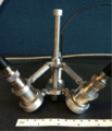
| − | + | Image:EVDGUI2.png|Photograph of the first prototype | |
</gallery> | </gallery> | ||
Latest revision as of 16:58, 23 June 2014
Home < 2014 Summer Project Week:Ventriculostomy Guidance Transcranial UltrasoundKey Investigators
- Jason White
- Kirby Vosburgh
- Can Meral
- Alex Golby
- Students: Abhishek Mundra, Michael Persaud, Aaron Silva
Project Description
Objective
- A ventriculostomy is often performed to relieve symptoms of emergent hydrocephalus. This involves the placement of an external ventricular drain (EVD) into the cerebral ventricles to remove excess cerebrospinal fluid. Free-handed EVD cannulation results in high rates of misplacement (~50%), leading to an increased risk of iatrogenic complications. Extant technical approaches to improve ventriculostomy guidance are either too complex or inaccurate. We have investigated the possibility of a novel device to guide EVD placement using transcranial ultrasound. The device uses three specifically aligned transducers delivering pulse-echo 0.5-MHz ultrasound through the skull bone to detect and localize the targeted ventricle. It also incorporates a cannula guide that is registered with the ultrasound FOV to integrate guidance with surgery.
Approach, Plan
- We wish to integrate and register the three A-mode US signals of the prototype device with MR or CT data during the investigation phase to assess the performance of the method. Prior work (Jason White, Alex Golby) in using 3DSlicer to combine pre-opertative images with intra-operative transcranial ultrasound for tracking brain shift will be built upon.


