Difference between revisions of "2016 Winter Project Week/Projects/BatchImageAnalysis"
From NAMIC Wiki
RandyGollub (talk | contribs) |
|||
| (12 intermediate revisions by 3 users not shown) | |||
| Line 1: | Line 1: | ||
__NOTOC__ | __NOTOC__ | ||
<gallery> | <gallery> | ||
| − | Image:PW-MIT2016.png|[[2016_Winter_Project_Week#Projects|Projects List]] | + | Image:PW-MIT2016.png|link=2016_Winter_Project_Week#Projects|[[2016_Winter_Project_Week#Projects|Projects List]] |
| + | <!-- Use the "Upload file" link on the left and then add a line to this list like "File:MyAlgorithmScreenshot.png" --> | ||
</gallery> | </gallery> | ||
| + | |||
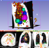
| + | [[File:LungCT-3DSIFT.png|200px|thumb|left|3D SIFT Lung Features]] | ||
==Key Investigators== | ==Key Investigators== | ||
| + | <!-- Add a bulleted list of investigators and their institutions here --> | ||
| + | |||
* Kalli Retzepi (MGH) | * Kalli Retzepi (MGH) | ||
* Yangming Ou (MGH) | * Yangming Ou (MGH) | ||
| − | * Matt Toews ( | + | * Matt Toews (ETS) |
| + | * Lilla Zollei (MGH) | ||
| + | * Lauren O'Donnell (BWH) | ||
* Steve Pieper (BWH) | * Steve Pieper (BWH) | ||
* Sandy Wells (BWH) | * Sandy Wells (BWH) | ||
| Line 13: | Line 20: | ||
==Project Description== | ==Project Description== | ||
| − | + | {| class="wikitable" | |
| − | + | ! style="text-align: left; width:27%" | Objective | |
| − | + | ! style="text-align: left; width:27%" | Approach and Plan | |
| − | + | ! style="text-align: left; width:27%" | Progress and Next Steps | |
| − | + | |- style="vertical-align:top;" | |
| − | + | | | |
| − | < | + | <!-- Objective bullet points --> |
| − | * | + | * Run feature detection code over a collection of medical images pulled from PACS |
| − | + | * Investigate a collection of ADC maps of neonates (diffusion MR) | |
| − | < | + | * Patients labeled with age and health status (normal, mildly abnormal, severely abnormal) |
| − | < | + | * Use 3D SIFT code to see if health status can be detected in images |
| − | * | + | * (if time) try text analysis of radiology reports |
| − | + | | | |
| − | + | <!-- Add a bulleted list of key points --> | |
| + | * Use deidentified cohort of neonate images collected from MGH | ||
| + | * Install data and software on AWS, try StarCluster | ||
| + | * Explore visualization options | ||
| + | * (if time) integrate image features with analysis of radiology report text | ||
| + | | | ||
| + | <!-- Fill this out at the end of Project Week; describe what you did this week and what you plan to do next --> | ||
| + | |||
| + | Algorithm | ||
| + | |||
| + | * feature extraction (20 seconds per image) | ||
| + | |||
| + | * Feature matching O(log N) indexing (< 1 second per image) | ||
| + | |||
| + | * 3D SIFT-Rank code (Windows, Linux, Max) and read me | ||
| + | http://www.matthewtoews.com/fba/featExtract1.5.zip | ||
| + | |||
| + | Result | ||
| + | Baseline HIE classification rate: 73%, leave-one-out moderate vs normal. | ||
| + | |||
| + | |||
| + | Data | ||
| + | 231 subjects, Apparent Diffusion Coefficient (ADC) images. | ||
| + | |||
| + | |} | ||
| + | |||
| + | ==Features Extracted in ADC MRI Volume == | ||
| + | |||
| + | http://wiki.na-mic.org/Wiki/images/b/b2/Image_%282%29.png | ||
Latest revision as of 15:54, 8 January 2016
Home < 2016 Winter Project Week < Projects < BatchImageAnalysisKey Investigators
- Kalli Retzepi (MGH)
- Yangming Ou (MGH)
- Matt Toews (ETS)
- Lilla Zollei (MGH)
- Lauren O'Donnell (BWH)
- Steve Pieper (BWH)
- Sandy Wells (BWH)
- Randy Gollub (MGH)
Project Description
| Objective | Approach and Plan | Progress and Next Steps |
|---|---|---|
|
|
Algorithm
http://www.matthewtoews.com/fba/featExtract1.5.zip Result Baseline HIE classification rate: 73%, leave-one-out moderate vs normal.
|
Features Extracted in ADC MRI Volume


