Difference between revisions of "Projects:ClusteringOfAnatomicallyDistinctFiberTracts"
| Line 21: | Line 21: | ||
[[Image:Th_corpus-SM_020001.png|[[Image:Th_corpus-SM_020001.png|Image:Th_corpus-SM_020001.png]]]] [[Image:Th_corpus-SM_140001.png|[[Image:Th_corpus-SM_140001.png|Image:Th_corpus-SM_140001.png]]]] [[Image:Th_corpus-SM_150001.png|[[Image:Th_corpus-SM_150001.png|Image:Th_corpus-SM_150001.png]]]] | [[Image:Th_corpus-SM_020001.png|[[Image:Th_corpus-SM_020001.png|Image:Th_corpus-SM_020001.png]]]] [[Image:Th_corpus-SM_140001.png|[[Image:Th_corpus-SM_140001.png|Image:Th_corpus-SM_140001.png]]]] [[Image:Th_corpus-SM_150001.png|[[Image:Th_corpus-SM_150001.png|Image:Th_corpus-SM_150001.png]]]] | ||
| + | ''References'' | ||
'''Key Investigators''' | '''Key Investigators''' | ||
Revision as of 13:00, 4 September 2007
Home < Projects:ClusteringOfAnatomicallyDistinctFiberTractsClustering Of Anatomically Distinct Fiber Tracts
Back to NA-MIC_Collaborations, MIT Algorithms
Objective
Use fiber clustering in multiple subjects to create a model ("atlas") of tractography clusters. Perform automatic segmentation of novel tractography datasets (brains) by application of this atlas. Also transfer anatomical information via cluster labels in the atlas.
Progress
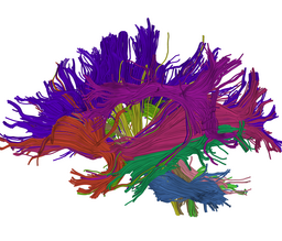
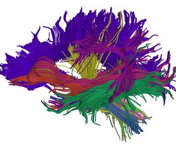
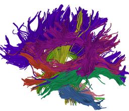
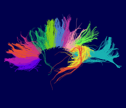
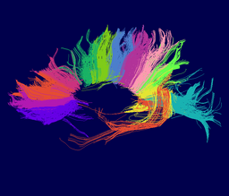
Atlas creation and automatic labeling has been performed in high-quality DTI datasets from Susumu Mori. Images showing example segmentation results are below. Work is underway to apply this atlas to segment additional datasets to define regions of interest that may be used in the study of schizophrenia.
Example Results
Selected anatomical regions, automatically labeled using the cluster atlas in 3 subjects.
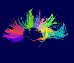
Subdivisions of the corpus callosum, labeled using the cluster atlas in 3 subjects.
References
Key Investigators
- MIT, Lauren O'Donnell
- BWH, Carl-Fredrik Westin
- BWH, Marek Kubicki
- BWH, Martha Shenton
Links
