Difference between revisions of "NA-MIC Brains Collaboration"
From NAMIC Wiki
| Line 33: | Line 33: | ||
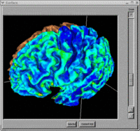
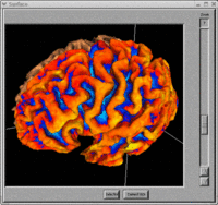
Image:BRAINS-Tissue-Clasification.gif|BRAINS Continuous Tissue Classification | Image:BRAINS-Tissue-Clasification.gif|BRAINS Continuous Tissue Classification | ||
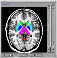
Image:ANN-Axial.gif|Automated segmentation of Subcortical nuclei using Neural Networks | Image:ANN-Axial.gif|Automated segmentation of Subcortical nuclei using Neural Networks | ||
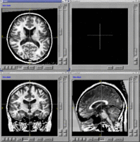
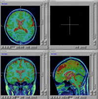
| − | Image:BRAINS-Image-Registration|BRAINS Image Registration using ITK Mutual Information | + | Image:BRAINS-Image-Registration.gif|BRAINS Image Registration using ITK Mutual Information |
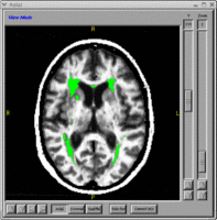
| + | Image:FLAIR-Lesion-Labels.gif|Lesion identification with FLAIR, T1 and T2 weighted images | ||
| + | Image:BRAINS-Talairach-Labels.gif|Talairach lobar labels | ||
| + | Image:GTRACT-Fiber-Tracking.gif|GTRACT DTI fiber tracking | ||
</gallery> | </gallery> | ||
Revision as of 00:56, 22 October 2007
Home < NA-MIC Brains CollaborationBack to NA-MIC_External_Collaborations
Contents
Abstract
This project is not an NCBC collaboration grant, but instead a Continued Development and Maintenance of Software grant. The intent of this application is to update the BRAINS image analysis software developed at the University of Iowa.
Grant #
1R01NS050568
Key Personnel
The collaborators include Vincent Magnotta, Hans Johnson, Jeremy Bockholt, and Nancy Andreasen.
Projects
There are three main thrusts of this application as outlined below.
Implement an Automated Brain Analysis Pipeline
Code Refactoring for Cross Platform Support
- Refactor Current Modules using ITK and VTK
- Build GUI using Slicer3 Interface
- Use Python as Scripting Language
Validation of Pipeline Using MIND MCIC Sample
- Evaluate MIND MCIC Human Phantom Sample Using Automated Pipeline
- Evaluate Differences between Patients with Schizophrenia and Normal Controls using Automated Pipeline
BRAINS Features
- BRAINS Gallery
Publications
- Powell S, Magnotta VA, Johnson H, Jammalamadaka VK, Pierson R, Andreasen NC. Registration and machine learning-based automated segmentation of subcortical and cerebellar brain structures. Neuroimage. 2007 Aug 22; [Epub ahead of print].
