Difference between revisions of "Events:HST-563-March2008"
| Line 34: | Line 34: | ||
===Reccommended exersie before the class=== | ===Reccommended exersie before the class=== | ||
| − | + | If you are interested in learing how we process pre-operative images in preparation for MRI-guided therapy, dowload free software slicer following the instruction in the "Slicer 101" page below. | |
| − | |||
| − | |||
| − | |||
http://wiki.na-mic.org/Wiki/index.php/Slicer:Workshops:User_Training_101 | http://wiki.na-mic.org/Wiki/index.php/Slicer:Workshops:User_Training_101 | ||
| − | + | The Slicer 101 tutorial will let you learn | |
| − | |||
| − | |||
*Data Loading and Visualization | *Data Loading and Visualization | ||
*Data Saving | *Data Saving | ||
| Line 55: | Line 50: | ||
*Dicom to Nrrd Conversion | *Dicom to Nrrd Conversion | ||
*Functional Magnetic Resonace Imaging Analysis | *Functional Magnetic Resonace Imaging Analysis | ||
| − | |||
| − | |||
| − | |||
==On the day of the lab....== | ==On the day of the lab....== | ||
Revision as of 21:26, 14 March 2008
Home < Events:HST-563-March2008Contents
Lab date/time/location
- March 19, 2008
- [Direction http://www.spl.harvard.edu/pages/Directions]
Background:
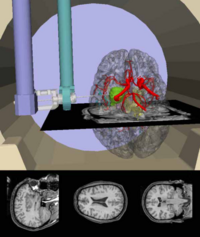
In comparison to other medical imaging modalities, magnetic resonance imaging (MRI) has clear advantages in its volumetric scanning capabilities, tissue discrimination and the detailed delineation of anatomic features. Additionally, MR imaging has unique potential capabilities for functional physiologic imaging and temperature mapping. These features of MR imaging motivated the development of intraoperative MR (IMRI) imaging for guidance of biopsy, thermal ablation and other surgical procedures. The major application of IMRI has been neurosurgical cases, including biopsy, drainage and tumor ablation. Other applications were liver biopsy and sinus endoscopic surgery.
There are many benefits in using pre-operatively obtained MRI as part of an analysis of imagery from IMRI. For example, IMRI has some limitations in its imaging capability in comparison to pre-operative MRI from conventional diagnostic MR scanner, since requirements for interventional use (for example, the use of surface coils for good access) have some impact on the imaging capability. After performing a registration that determines the proper spatial relationship between the pre-operative MRI and IMRI, one could compare and determine whether changes in tissue structure have occurred. For instance, this would be extremely useful in evaluating the extent of tumor.
We can further appreciate the benefit of pre-operative image, if we can co-register imagery from other modalities than MRI. Currently, we can register computed tomography (CT), T1- and T2-weighted MRI: magnetic resonance angiography (MRA), single photon emission computed tomography (SPECT). Incorpolatin of these images can provide information that can bot be deduced from regular MRI. In general, utilizing pre- and intra-operative image registration would allow a surgeon to more precisely identify and avoid critical structures and more accurately locate pathological tissues during a procedure.
In this paper, we will report our approach to register pre-operative image to intra-operative MRI and use co-registered images to navigate neurosurgeries. We will first outline the registration method through maximization of mutual information. Then, the comprehensive accuracy study of the registration and clinical application of the method are introduced. In the clinical application section, we will introduce our engineering and computational setup to achieve online and near real-time registration and navigation in an inerventional MRI scanner (Signa SP, General Electric Medical Systems, Milwaukee, WI). An example scenario
Recommended reading before the class
http://www.spl.harvard.edu/pages/Special:PubDB_View?dspaceid=1225
http://www.spl.harvard.edu/pages/Special:PubDB_View?dspaceid=294
http://www.spl.harvard.edu/pages/Special:PubDB_View?dspaceid=1244
http://www.spl.harvard.edu/pages/Special:PubDB_View?dspaceid=1247
Reccommended exersie before the class
If you are interested in learing how we process pre-operative images in preparation for MRI-guided therapy, dowload free software slicer following the instruction in the "Slicer 101" page below. http://wiki.na-mic.org/Wiki/index.php/Slicer:Workshops:User_Training_101
The Slicer 101 tutorial will let you learn
- Data Loading and Visualization
- Data Saving
- Manual Segmentation
- Level-Set Segmentation
- Automatic Brain Segmentation
- Registration
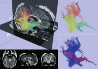
- Diffusion Tensor Imaging Analysis
- Nrrd File Format
- Dicom to Nrrd Conversion
- Functional Magnetic Resonace Imaging Analysis
On the day of the lab....
Introdcution
Live demonstration
Homework problems
- What is the benefit of medical images for guiding and navigating surgery?
- What are the technology involved in Image guided Therapy? List and briefly describe them in the order of workflow?
- What is the patient-to-image registration? What kind of mathematical process is involved in the patient-to-image registration?
- What is Target Registration Error, Fiducial Registration Error, and Fiducial Localization error?
- What is the difference between pre-operative image guided therapy and intra-operative image guided therapy?
- What is the benefit of MRI-guided Intra-operative image guided therapy?
- How would you solve the problem that intra-operative MRI has inherently less quality than pre-operative images?
- What are the main clinical application in MRI-guided therapy?
- From PubMed, find three interesting paper on MRI-guided therapy and summerize them.
- What do you think is unchallenged clinical target in MRI-guided therapy?

