Difference between revisions of "2010 Winter Project Week Spine Segmentation Module in Slicer3"
Sylvainjaume (talk | contribs) (Create page for 2010 Winter Week project about Spine Segmentation from MRI data) |
Sylvainjaume (talk | contribs) (Add screenshots) |
||
| Line 2: | Line 2: | ||
<gallery> | <gallery> | ||
Image:PW-SLC2010.png|[[2010_Winter_Project_Week#Projects|Projects List]] | Image:PW-SLC2010.png|[[2010_Winter_Project_Week#Projects|Projects List]] | ||
| − | Image: | + | Image:spine-segmentation-jaume2009.png|MRI image of a patient with disc herniation |
| − | Image: | + | Image:spine-segmentation-slicer2009.png|Segmentation of the herniated disc and the cerebro-spinal fluid using Slicer 3.5 |
</gallery> | </gallery> | ||
Revision as of 18:59, 4 December 2009
Home < 2010 Winter Project Week Spine Segmentation Module in Slicer3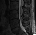
MRI image of a patient with disc herniation
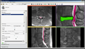
Segmentation of the herniated disc and the cerebro-spinal fluid using Slicer 3.5


Key Investigators
- Heidelberg University: Martin Loepprich
- MIT: Sylvain Jaume, Polina Golland
- BWH: Ron Kikinis, Steve Pieper
Objective
We are developing a Slicer module to segment the region within the thecal sac in MRI images of the spine. Our objective is to provide a segmentation and visualization tool to improve the treatment of disc herniation. The structures of interests are the cerebro-spinal fluid (CSF), the discs, the vertebrae and the spinal nerves. The main challenge is to perform the segmentation in a fully automated way.
Approach, Plan
Our approach is to design a pattern recognition and analyze the statistics of the automated recognition to learn the optimal parameters. Our plan for the project week is to integrate our code into Slicer 3.5.
Progress
The algorithm is currently being implemented using ITK and VTK.
References
- Segmentation using Slicer 3.5, EMSegment module