Difference between revisions of "2011 Winter Project Week:StenosisDetector"
From NAMIC Wiki
| Line 2: | Line 2: | ||
<gallery> | <gallery> | ||
Image:PW-SLC2011.png|[[2011_Winter_Project_Week#Projects|Projects List]] | Image:PW-SLC2011.png|[[2011_Winter_Project_Week#Projects|Projects List]] | ||
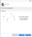
| + | Image:step1.png|StensosisDetector Step1 | ||
</gallery> | </gallery> | ||
| Line 14: | Line 15: | ||
<h3>Objective</h3> | <h3>Objective</h3> | ||
| − | We are developing a stenosis detector based on VMTK in Slicer 4. The goal is to be able to visualize stenosis after a vessel segmentation. | + | We are developing a stenosis detector based on VMTK in Slicer 4. The goal is to be able to visualize stenosis after a vessel segmentation using a wizard-based interface. |
Revision as of 19:54, 16 December 2010
Home < 2011 Winter Project Week:StenosisDetectorKey Investigators
- University of Heidelberg: Suares Tamekue
- UPenn: Daniel Haehn
- Mario Negri Institute, Italy: Luca Antiga
- SPL: Ron Kikinis
Objective
We are developing a stenosis detector based on VMTK in Slicer 4. The goal is to be able to visualize stenosis after a vessel segmentation using a wizard-based interface.
Approach, Plan
Our approach for developing the stenosis detector is: first vessel enhancement, level-set segmentation, network extraction and then quantification and visualization of stenosis.
The tool will be evaluated on datasets.
Progress
A prototyp of the graphical user interface has been designed and implemented.
Delivery Mechanism
This work will be delivered to the NA-MIC Kit as a (please select the appropriate options by noting YES against them below)
- ITK Module
- Slicer Module
- Built-in
- Extension -- commandline
- Extension -- loadable [X]
- Other (Please specify)
References
- Antiga L, Piccinelli M, Botti L, Ene-Iordache B, Remuzzi A and Steinman DA. An image-based modeling framework for patient-specific computational hemodynamics. Medical and Biological Engineering and Computing, 46: 1097-1112, Nov 2008.
- D. Hähn. Integration of the vascular modeling toolkit in 3d slicer. SPL, 04 2009. Available online at http://www.spl.harvard.edu/publications/item/view/1728.
- D. Hähn. Centerline Extraction of Coronary Arteries in 3D Slicer using VMTK based Tools. Master's Thesis. Department of Medical Informatics, University of Heidelberg, Germany. Feb 2010.
- Piccinelli M, Veneziani A, Steinman DA, Remuzzi A, Antiga L (2009) A framework for geometric analysis of vascular structures: applications to cerebral aneurysms. IEEE Trans Med Imaging. In press.

