Difference between revisions of "DBP3:Utah:AutoWallSeg"
From NAMIC Wiki
(Created page with '=Automatic Segmentation= ==Automatic Wall Segmentation from GA Tech== GA Tech produced a slicer module to automatically segment the left atrial wall, given the original data AN…') |
|||
| Line 4: | Line 4: | ||
GA Tech produced a slicer module to automatically segment the left atrial wall, given the original data AND the endo (blood pool) segmentation. Below is some evaluation of those segmentations. | GA Tech produced a slicer module to automatically segment the left atrial wall, given the original data AND the endo (blood pool) segmentation. Below is some evaluation of those segmentations. | ||
| + | |||
| + | {| class="wikitable" | ||
| + | |- | ||
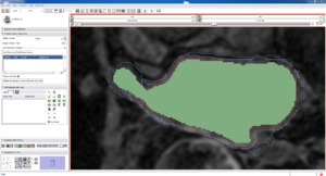
| + | ! Auto, Endo, and Manaul Wall (01 3mo s5) | ||
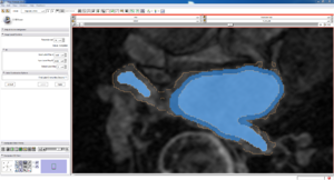
| + | ! Auto, Endo, and Manual Wall (01 3mo s4) | ||
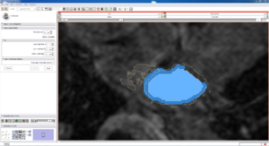
| + | ! Auto, Endo, and Manual Wall (01 3mo s1) | ||
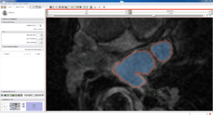
| + | ! Auto, Endo, and 4 Pixel Dilation from Endo (01 3mo s1) | ||
| + | |- | ||
| + | | [[File:01 3mo s5 Auto Endo Manual slice.png|thumb]] | ||
| + | | [[File:01 3mo s4 Auto Endo Manual slice.png|thumb]] | ||
| + | | [[File:01 3mo s1 Auto Endo Manual slice.png|thumb]] | ||
| + | | [[File:01 3mo s1 Auto Endo 4PixDilate slice.png|thumb]] | ||
| + | |- | ||
| + | | This image shows how the automatic wall segmentation creates a complete surface around the endo - while a manual segmentor would cut off the wall around the veins (although the location this cutoff decision is arbitrary). | ||
| + | | This image shows where the automatic segmentation surrounds a vein, while the manual segmentor decided to not surround the vein. | ||
| + | | This image shows some island artifacts that may be the result of the process being done in 3D (leaking from the slice above) - note that the data below the islands is lighter. | ||
| + | | Here we show a 4 pixel dilation from the endo compared to the automatic segmentation. This also image shows more islands. | ||
| + | |- | ||
| + | |} | ||
Revision as of 00:21, 22 April 2011
Home < DBP3:Utah:AutoWallSegAutomatic Segmentation
Automatic Wall Segmentation from GA Tech
GA Tech produced a slicer module to automatically segment the left atrial wall, given the original data AND the endo (blood pool) segmentation. Below is some evaluation of those segmentations.
| Auto, Endo, and Manaul Wall (01 3mo s5) | Auto, Endo, and Manual Wall (01 3mo s4) | Auto, Endo, and Manual Wall (01 3mo s1) | Auto, Endo, and 4 Pixel Dilation from Endo (01 3mo s1) |
|---|---|---|---|
| This image shows how the automatic wall segmentation creates a complete surface around the endo - while a manual segmentor would cut off the wall around the veins (although the location this cutoff decision is arbitrary). | This image shows where the automatic segmentation surrounds a vein, while the manual segmentor decided to not surround the vein. | This image shows some island artifacts that may be the result of the process being done in 3D (leaking from the slice above) - note that the data below the islands is lighter. | Here we show a 4 pixel dilation from the endo compared to the automatic segmentation. This also image shows more islands. |


