Difference between revisions of "Projects:TubularSurfaceSegmentation"
| Line 1: | Line 1: | ||
| − | Back to [[NA-MIC_Internal_Collaborations:StructuralImageAnalysis|NA-MIC Collaborations]], [[Algorithm: | + | Back to [[NA-MIC_Internal_Collaborations:StructuralImageAnalysis|NA-MIC Collaborations]], [[Algorithm:Stony Brook|Stony Brook University Algorithms]], [[Engineering:Kitware|Kitware Engineering]] |
__NOTOC__ | __NOTOC__ | ||
= Tubular Surface Segmentation Framework = | = Tubular Surface Segmentation Framework = | ||
Latest revision as of 01:02, 16 November 2013
Home < Projects:TubularSurfaceSegmentationBack to NA-MIC Collaborations, Stony Brook University Algorithms, Kitware Engineering
Tubular Surface Segmentation Framework
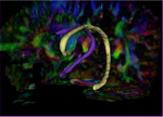
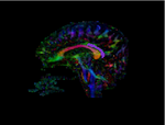
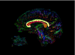
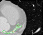
This is a new model for tubular surfaces that transforms the problem of detecting a surface in 3D space, to detecting a curve in 4D space. This model affords computation efficiency and stability (by the use of the Sobolev norm) and we have successfully tested its application in segmenting the Cingulum Bundle from DW-MRI of the brain as well as vessel segmentation from CT cardiac data.
Description
We have proposed a new model for tubular surfaces that represents a tubular surface as a center-line with a radius function associated with every point of the center-line. This transforms the problem of detecting a surface in 3D space, to detecting a curve in 4D space. Besides allowing us to impose a "soft" tubular shape prior, this also leads to computational efficiency over conventional surface segmentation approaches. We have also developed the moving end points implementation of this framework wherein the required input is only a few points in the interior of the structure of interest. This yields the additional advantage that the framework simultaneously returns both the 3D segmentation and the 3D skeleton of the structure eliminating the need for a priori knowledge of end points, and an expensive skeletonization step. The framework is applicable to different tubular anatomical structures in the body. We have so far applied it successfully to the Cingulum Bundle, and blood vessels.
Some Results
Project Status
- Algorithm successfully implemented and tested in MATLAB for Cingulum Bundle (as well as vessel segmentation).
Key Investigators
- Georgia Tech Algorithms: Vandana Mohan, Allen Tannenbaum
- Kitware Engineering: Luis Ibanez
Publications
In Press
- Mohan V., Sundaramoorthi G., Tannenbaum A. Tubular Surface Segmentation for Extracting Anatomical Structures from Medical Imagery. IEEE Transactions in Medical Imaging (in review). IEEE Transactions on Medical Imaging (2010) (see here).
Project Week Results: June 2009


