Difference between revisions of "Project Week 25/Slicer and 3D Printing"
From NAMIC Wiki
| Line 43: | Line 43: | ||
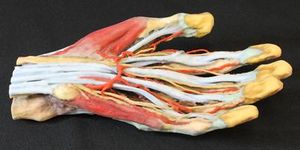
[[File:Nayra1.jpg|thumb|left|3D printed model (hand colored)]] | [[File:Nayra1.jpg|thumb|left|3D printed model (hand colored)]] | ||
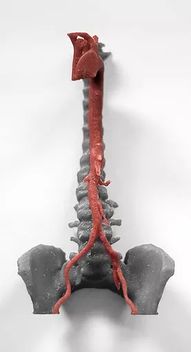
[[File:Nayra2.jpg|thumb|left|3D print of a human aorta]] | [[File:Nayra2.jpg|thumb|left|3D print of a human aorta]] | ||
| − | [[File:Marilola4.png|thumb|[[https://mt4sd.ulpgc.es/en/]]]] | + | [[File:Marilola4.png|thumb|left|[[https://mt4sd.ulpgc.es/en/]]]] |
==Background and References== | ==Background and References== | ||
<!-- Use this space for information that may help people better understand your project, like links to papers, source code, or data --> | <!-- Use this space for information that may help people better understand your project, like links to papers, source code, or data --> | ||
Revision as of 09:28, 15 June 2017
Home < Project Week 25 < Slicer and 3D Printing
Back to Projects List
Key Investigators
- Juan Ruiz Alzola (Universidad de Las Palmas de Gran Canaria, Spain)
- Michael Halle (Harvard University)
Project Description
From DICOM data to a 3D print of anatomical models for training: anatomy classes and/or surgical planning
| Objective | Approach and Plan | Progress and Next Steps |
|---|---|---|
|
|
Illustrations

[[1]]

