Difference between revisions of "2008 Winter Project Week:fmri eeg analysis"
From NAMIC Wiki
| Line 28: | Line 28: | ||
<h1>Progress</h1> | <h1>Progress</h1> | ||
| − | The SCIrun environment was compiled on our Ubuntu laptop. | + | The SCIrun environment was compiled on our Ubuntu laptop. This will enable us to create |
| + | volume meshes, store eeg electrode locations and calculate electric field streamlines | ||
| + | in our brain data. | ||
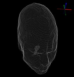
[[Image:scirunhead1.jpg|256px]] | [[Image:scirunhead1.jpg|256px]] | ||
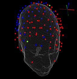
[[Image:scirunhead2.jpg|256px]] | [[Image:scirunhead2.jpg|256px]] | ||
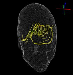
| − | [[Image:scirunhead3.jpg]] | + | [[Image:scirunhead3.jpg|256px]] |
Data currently acquired: | Data currently acquired: | ||
Revision as of 18:30, 11 January 2008
Home < 2008 Winter Project Week:fmri eeg analysis Return to 2008_Winter_Project_Week |
Key Investigators
- BWH: Padma Sundaram, Darren Orbach
Objective
We are interested in visualizing electromagnetic activity in the brain using MR imaging with concurrent EEG. We are particularly interested in inspecting epileptic foci and sleeping individuals.
Approach, Plan
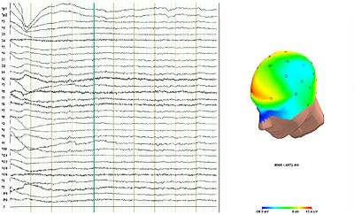
We would like to analyze the fMRI data within the slicer framework and also use SCIRun to analyze the EEG and visualize the bioelectric fields.
Progress
The SCIrun environment was compiled on our Ubuntu laptop. This will enable us to create volume meshes, store eeg electrode locations and calculate electric field streamlines in our brain data.
Data currently acquired:
- Rest-state fMRI and concurrent EEG (normal healthy volunteer)
- EEG of a sleeping individual (no MRI yet, but data covers 8 pm to 10 am and all sleep stages)
References
- Direct detection of neuronal activity with MRI: fantasy, possibility, or reality?, Bandettini, N. Petridou and J. Bodurka, Appl. Magn. Reson. 29 (2005), pp. 65–88.
- Visual Analysis of Bioelectric Fields, Xavier Trioche, Rob MacLeod, Chris Johnson.
