Difference between revisions of "IGT:ToolKit/Prostate-Planning"
From NAMIC Wiki
| Line 3: | Line 3: | ||
Back to [[IGT:ToolKit|IGT:ToolKit]] | Back to [[IGT:ToolKit|IGT:ToolKit]] | ||
| − | = | + | =Prostate Therapy Planning Tutorial= |
==Overview== | ==Overview== | ||
| − | This tutorial | + | This tutorial gives an example of using Slicer3 for MRI-guided prostate interventions. Topics covered include: |
| − | * | + | * Clinical background on MRI-guided prostate interventions |
| − | + | * Non-rigid image registration using B-splines | |
| − | * | ||
| − | |||
==Tutorial Materials== | ==Tutorial Materials== | ||
| − | |||
| − | |||
| − | |||
| − | |||
| − | |||
| − | |||
| − | |||
| − | + | * Tutorial slides for the MRI-guided prostate interventions tutorial: [[Media:MRGuidedProstateInterventions.pdf| pdf]] (recommended) or [[Media:MRGuidedProstateInterventions.ppt | ppt]] | |
| − | + | * [[Media:NeurosurgicalPlanningTutorialData.zip | MRI-guided prostate interventions tutorial dataset]] - contains: | |
| − | + | ** Pre-operative high-quality MRI image | |
| − | + | ** Intra-operative MRI image | |
| − | |||
| − | |||
| − | *[[ | ||
==Software Installation Instructions== | ==Software Installation Instructions== | ||
| Line 36: | Line 24: | ||
* [http://www.spl.harvard.edu/~dpace Danielle Pace] | * [http://www.spl.harvard.edu/~dpace Danielle Pace] | ||
* [http://www.spl.harvard.edu/~noby Nobuhiko Hata] | * [http://www.spl.harvard.edu/~noby Nobuhiko Hata] | ||
| − | |||
| − | |||
| − | |||
| − | |||
| − | |||
| − | |||
| − | |||
| − | |||
Revision as of 20:34, 25 November 2008
Home < IGT:ToolKit < Prostate-PlanningBack to IGT:ToolKit
Contents
Prostate Therapy Planning Tutorial
Overview
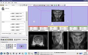
This tutorial gives an example of using Slicer3 for MRI-guided prostate interventions. Topics covered include:
- Clinical background on MRI-guided prostate interventions
- Non-rigid image registration using B-splines
Tutorial Materials
- Tutorial slides for the MRI-guided prostate interventions tutorial: pdf (recommended) or ppt
- MRI-guided prostate interventions tutorial dataset - contains:
- Pre-operative high-quality MRI image
- Intra-operative MRI image
Software Installation Instructions
Go to the Slicer3 Install site.
