Difference between revisions of "2009 Winter Project Week Tumor Microenvironment"
From NAMIC Wiki
| Line 2: | Line 2: | ||
|[[Image:NAMIC-SLC.jpg|thumb|320px|Return to [[2008_Winter_Project_Week]] ]] | |[[Image:NAMIC-SLC.jpg|thumb|320px|Return to [[2008_Winter_Project_Week]] ]] | ||
|[[Image:Tme_snapshot2.jpg|thumb|320px|]] | |[[Image:Tme_snapshot2.jpg|thumb|320px|]] | ||
| − | |[[Image: | + | |[[Image:snap3.jpg|thumb|320px|]] |
|} | |} | ||
Revision as of 16:58, 5 January 2009
Home < 2009 Winter Project Week Tumor Microenvironment Return to 2008_Winter_Project_Week |
Key Investigators
- The Ohio State University: Shantanu Singh, Raghu Machiraju
- GE Global Research: Jens Rittscher
- BWH: Michael Halle, Steve Pieper, Wendy Plesniak
Objective
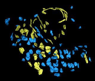
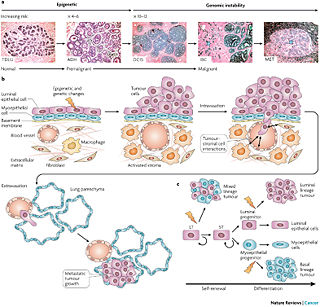
To implement a microscopy image analysis pipeline in Slicer3, specifically tailored for characterizing the tumor microenvironment as observed in the murine breast tumors.
Approach, Plan
Integrate existing segmentation, validation and labeling pipelines into the Slicer3 framework. Begin with level set methods of Mosaliganti et al. Then the tessellation based method of the same authors, time permitting. Explore what can be obtained with EMSegement of Slicer3. Compare and contrast all sets of results. Also, explore preliminary labeling.
Progress
Updated every day.

