Difference between revisions of "Algorithm:Utah2"
| Line 1: | Line 1: | ||
| − | Back to [[Algorithm:Main|NA-MIC Algorithms]] | + | Back to [[Algorithm:Main|NA-MIC Algorithms]] |
__NOTOC__ | __NOTOC__ | ||
= Overview of Utah 2 Algorithms (PI: Guido Gerig) = | = Overview of Utah 2 Algorithms (PI: Guido Gerig) = | ||
| Line 7: | Line 7: | ||
= Utah 2 Projects = | = Utah 2 Projects = | ||
| − | {| cellpadding="10" | + | {| cellpadding="10" style="text-align:left;" |
| style="width:15%" | [[Image:Cbg-dtiatlas-tracts.png|200px]] | | style="width:15%" | [[Image:Cbg-dtiatlas-tracts.png|200px]] | ||
Revision as of 19:05, 11 July 2009
Home < Algorithm:Utah2Back to NA-MIC Algorithms
Overview of Utah 2 Algorithms (PI: Guido Gerig)
At Utah, we are interested in a range of algorithms and solutions for the analysis of DTI and the segmentation of healthy brains and brains with lupus lesions from structural MRI.
Utah 2 Projects

|
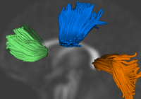
Group Analysis of DTI Fiber TractsAnalysis of populations of diffusion images typically requires time-consuming manual segmentation of structures of interest to obtain correspondance for statistics. This project uses non-rigid registration of DTI images to produce a common coordinate system for hypothesis testing of diffusion properties. More... New: Casey B. Goodlett, P. Thomas Fletcher, John H. Gilmore, Guido Gerig. Group Analysis of DTI Fiber Tract Statistics with Application to Neurodevelopment. NeuroImage 45 (1) Supp. 1, 2009. p. S133-S142. pubdb |

|
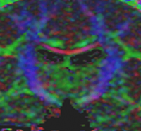
Influence of Imaging Noise on DTI StatisticsClinical acquisition of diffusion weighted images with high signal to noise ratio remains a challenge. The goal of this project is to understand the impact of MR noise on quantiative statistics of diffusion properties such as anisotropy measures, trace, etc. More... New: Casey Goodlett, P. Thomas Fletcher, Weili Lin, Guido Gerig. Quantification of measurement error in DTI: Theoretical predictions and validation, MICCAI 2007. |

|
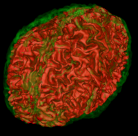
Atlas Based Brain SegmentationAutomatic segmentation can be performed reliably using priors from brain atlases and an image generative model. We have developed a tool that provides an automatic segmentation pipeline in a modular framework. More... |

|
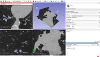
Lesion SegmentationQuantification, analysis and display of brain pathology such as white matter lesions as observed in MRI is important for diagnosis, monitoring of disease progression, improved understanding of pathological processes and for developing new therapies. More... |