Difference between revisions of "DBP2:UNC:Local Cortical Thickness Pipeline"
| Line 139: | Line 139: | ||
[http://insight-journal.org/midas/community/view/24 Data of 2 autistic children and 2 normal controls] (male, female) scanned at 2 years with follow up at 4 years from a 1.5T Siemens scanner. Files include structural data, tissue segmentation label map and subcortical structures segmentation. | [http://insight-journal.org/midas/community/view/24 Data of 2 autistic children and 2 normal controls] (male, female) scanned at 2 years with follow up at 4 years from a 1.5T Siemens scanner. Files include structural data, tissue segmentation label map and subcortical structures segmentation. | ||
| + | ==== Tutorials ==== | ||
| + | • '''GAMBIT tutorial''' : end-to-end Slicer3 module to perform group-wise automatic mesh-based analysis of cortical thickness [[Media:GAMBIT_TutorialContestSummer2010.ppt| [ppt]]][[Media:GAMBIT_TutorialContestSummer2010.pdf| [pdf]]] | ||
== Pipeline validation == | == Pipeline validation == | ||
Revision as of 21:09, 26 August 2010
Home < DBP2:UNC:Local Cortical Thickness PipelineBack to UNC Cortical Thickness Roadmap
Objective
We would like to create an end-to-end application within Slicer3 allowing group-wise automatic mesh-based analysis of cortical thickness as well as other surface measurements (surface area...)
This page describes the related pipeline with its basic components, as well as its validation.
Pipeline overview
A Slicer3 high-level module for group-wise cortical thickness analysis has been developed: GAMBIT (Group-wise Automatic Mesh Based analysis of cortIcal Thickness)
Input: CSV file containing RAW images (T1-weighted, T2-weighted, PD-weighted images)
- 1. Individual pipeline
- 1.1. Tissue segmentation
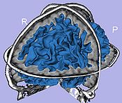
- Probabilistic atlas-based automatic tissue segmentation via an Expectation-Maximization scheme
- Tool: itkEMS (UNC Slicer3 external module)
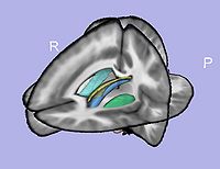
- 1.2. Atlas-based ROI segmentation: subcortical structures, lateral ventricles, parcellation
- 1.2.1. Skull stripping using previously computed tissue segmentation label image
- Tool: SegPostProcess (UNC Slicer3 external module)
- 1.2.2. T1-weighted atlas deformable registration
- B-spline pipeline registration
- Tool: RegisterImages (Slicer3 module)
- 1.2.3. Applying transformations to the structures
- Tool: ResampleVolume2 (Slicer3 module)
- 1.2.1. Skull stripping using previously computed tissue segmentation label image
- 1.3. White matter map creation
- Brainstem and cerebellum extraction
- Adding subcortical structures except amygdala and hippocampus
- Tool: ImageMath (UNC Slicer3 external module)
- 1.4. Cortical thickness computation
- Asymmetric cortical thickness
- Tool: UNCCortThick(UNC Slicer3 external module)
- 1.5. White matter map post-processing
- Largest component computation
- Smoothing: Level set smoothing or weighted average filter
- Connectivity enforcement (6-connectivity)
- White matter filling
- Tool: WMSegPostProcess (UNC Slicer3 external module)
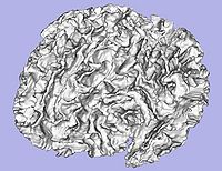
- 1.6. Genus zero white matter map image and surface creation
- Tool: GenusZeroImageFilter (UNC Slicer3 external module)
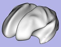
- 1.7. White matter surface inflation
- Iterative smoothing using relaxation operator (considering average vertex) and L2 norm of the mean curvature as a stopping criterion
- Iteration stopped if vertices that have too high curvature (some extremities)
- Tool: MeshInflation (UNC Slicer3 external module)
- 1.7 bis(Optional). White matter image fixing if necessary
- Correction of the white matter map image (corresponding to vertices that have high curvature) with connectivity enforcement
- Tool: FixImage (UNC Slicer3 external module)
- Go back to step 1.6
- 1.8. Sulcal depth
- Sulcal depth computation using genus-zero surface and inflated one
- Tool: MeshMath (UNC module)
- 1.9. Surface area computation
- Lobar surface area measurement on smoothed genus-zero surface
- Tool: MeshMath (UNC module)
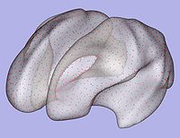
- 1.10. Particles initialization for cortical correspondence
- Initializing particles on inflated genus-zero surface using 98-lobe parcellation map and genus zero surface
- Tool: ParticleInitializer (UNC Slicer3 external module)
- 1.1. Tissue segmentation
- 2. Particle-based shape correspondence
- Correspondence on inflated surfaces using particle system
- 2.1. Preprocessing
- Distance maps creation from inflated genus-zero surfaces with slight gaussian blurring
- Tool: ParticleCorrespondencePreProcessing (UNC Slicer3 external module)
- 2.2. Correspondence optimization
- Particle-based shape correspondence optimization (using sulcal depth) with Procrustes alignement
- Tool: ShapeWorksRun (Utah Slicer3 external module)
- 2.3. Postprocessing
- Re-meshing using template
- Tool: ParticleCorrespondencePostProcessing (UNC Slicer3 external module)
- 2.4. Cortical thickness interpolation
- Cortical thickness interpolation on surface in correspondence
- Tool: MeshMath (UNC module)
- 3. Group statistical analysis
- Tool: QDEC Slicer module or StatNonParamPDM
Download
All the tools used in the current pipeline are Slicer3 modules, some of them being UNC external modules. The user can thus compute an individual regional cortical thickness analysis by running the 'GAMBIT' module, either within Slicer3 or as a command line.
GAMBIT download
Source code, executables and tutorial are available on NITRC
Complementary downloads
Brain atlases
Four brain atlases are available on MIDAS and on NITRC:
Pediatric MRI Brain data
Data of 2 autistic children and 2 normal controls (male, female) scanned at 2 years with follow up at 4 years from a 1.5T Siemens scanner. Files include structural data, tissue segmentation label map and subcortical structures segmentation.
Tutorials
• GAMBIT tutorial : end-to-end Slicer3 module to perform group-wise automatic mesh-based analysis of cortical thickness [ppt] [pdf]
Pipeline validation
Analysis on a small pediatric dataset
Tests will be computed on a small pediatric dataset which includes 2 year-old and 4 year-old cases.
- 16 autistic cases
- 1 developmental delay
- 3 normal control
Comparison to state of the art
We would like to compare our pipeline with FreeSurfer. We will thus perform a regional statistical analysis using Pearson's correlation coefficient on an adult dataset (FreeSurfer's publicly available tutorial dataset) including 40 cases.
Planning
Done
- Workflow for group analysis (Slicer3 external module using BatchMake):
- Development of UNC Slicer3 modules
- Modules applied on small pediatric dataset from the Autism study
- Pediatric and adult brain atlases available to the community via MIDAS
- T1-weighted atlas
- Tissue segmentation probability maps
- Subcortical structures probability maps
- LBinary mask images
- GAMBIT available to the community via NITRC: executables (UNC external modules for Slicer3) and tutorial dataset
- Tutorial with application example on a small dataset
- GAMBIT source code (CVS) available to the community
In progress
- Step 1.7: Parameter exploration on autism dataset to improve inflation-fixing steps
- Step 2: Particle correspondence testing with pediatric surfaces (Meeting with Josh Cates at UNC - February 2010)
- New version of GAMBIT including quality control through MRML scene, and WM, GM models generation
- GAMBIT executable can be downloaded directly within Slicer3 using the extension wizard
References
- C. Vachet, H.C. Hazlett, M. Niethammer, I. Oguz, J.Cates, R. Whitaker, J. Piven, M. Styner, Mesh-based Local Cortical Thickness Framework, UNC Radiology Research Day 2010 abstract
- I. Oguz, M. Niethammer, J. Cates, R. Whitaker, T. Fletcher, C. Vachet, and M. Styner, Cortical Correspondence with Probabilistic Fiber Connectivity, Information Processing in Medical Imaging, IPMI 2009, LNCS, 21:651-63
- C. Vachet, H.C. Hazlett, M. Niethammer, I. Oguz, J.Cates, R. Whitaker, J. Piven, M. Styner, Mesh-based Local Cortical Thickness Framework, UNC Radiology Research Day 2009 abstract
- Oguz, I., Cates, J., Fletcher, T., Whitaker, R., Cool, D., Aylward, S., Styner, M., Cortical correspondence using entropy-based particle systems and local features, IEEE Symposium on Biomedical Imaging ISBI 2008. 1637– 1640
- J. Cates, P. Fletcher, M. Styner, H. Hazlett, R. Whitaker, Particle-based shape analysis of multi-object complexes, MICCAI 2008, 477-85
- Cates, J., Fletcher, P., Whitaker, R.: Entropy-based particle systems for shape correspondence. MFCA Workshop, MICCAI 2006, 90–99
