Difference between revisions of "Projects:RegistrationLibrary:RegLib C13"
From NAMIC Wiki
| Line 61: | Line 61: | ||
#repeat and create a similar mask for the MRI-T2. | #repeat and create a similar mask for the MRI-T2. | ||
#save results thus far | #save results thus far | ||
| − | *'''Phase III: | + | *'''Phase III: Affine Registration MR-CT''' |
| + | :this phase runs a linear registration first to better track progress. We then use this registration as input for the nonrigid registration in phase IV below. You may combine the two and include a BSpline registration phaser right away. | ||
#open the [http://wiki.slicer.org/slicerWiki/index.php/Documentation/4.1/Modules/BRAINSFit General Registration (BRAINS) module] | #open the [http://wiki.slicer.org/slicerWiki/index.php/Documentation/4.1/Modules/BRAINSFit General Registration (BRAINS) module] | ||
##''Fixed Image Volume'': CTpost | ##''Fixed Image Volume'': CTpost | ||
| Line 67: | Line 68: | ||
##Output Settings: | ##Output Settings: | ||
###''Slicer BSpline Transform": none | ###''Slicer BSpline Transform": none | ||
| − | ###''Slicer Linear Transform'': create new transform, rename to "Xf2_T2-CTpost_Affine | + | ###''Slicer Linear Transform'': create new transform, rename to "Xf2_T2-CTpost_Affine" |
| − | ###''Output Image Volume'': create new volume, rename to "T2_Xf2" (we use this for validation only) | + | ###''Output Image Volume'': create new volume, rename to "MRI-T2_Xf2" (we use this for validation only) |
| − | ##''Registration Phases'': check boxes for ''Rigid'', "Rigid+Scale", "Affine | + | ##''Registration Phases'': check boxes for ''Rigid'', "Rigid+Scale", "Affine" |
##''Main Parameters'': | ##''Main Parameters'': | ||
##''Mask Option'': select ''ROI'' button | ##''Mask Option'': select ''ROI'' button | ||
###''ROI Masking input fixed'': select "CTpost_label" created in phase II above | ###''ROI Masking input fixed'': select "CTpost_label" created in phase II above | ||
###''ROI Masking input moving'': select "MRI-T2-label.nrrd" created in phase II above | ###''ROI Masking input moving'': select "MRI-T2-label.nrrd" created in phase II above | ||
| + | ##Leave all other settings at default | ||
| + | ##click: Apply | ||
| + | #you should get a registration similar to the one shown in the results section below. | ||
| + | |||
| + | *'''Phase IV: Nonrigid Registration MR-CT''' | ||
| + | #open the [http://wiki.slicer.org/slicerWiki/index.php/Documentation/4.1/Modules/BRAINSFit General Registration (BRAINS) module] | ||
| + | ##''Fixed Image Volume'': CTpost | ||
| + | ##''Moving Image Volume'': MRI_T2 | ||
| + | ##Output Settings: | ||
| + | ###''Slicer BSpline Transform": create new transform, rename to "Xf3_T2-CTpost_BSpline" | ||
| + | ###''Slicer Linear Transform'': none | ||
| + | ###''Output Image Volume'': create new volume, rename to "MRI-T2_Xf3" | ||
| + | ##''Registration Phases'': check boxes for ''BSpline'' only | ||
| + | ##''Main Parameters'': | ||
| + | ###''Number Of Samples'': 200,000 | ||
| + | ###''B-Spline Grid Size'': 5,5,5 | ||
| + | ##''Mask Option'': select ''ROI'' button | ||
| + | ###''ROI Masking input fixed'': select " "T1_mask_ROIauto" or "T1_mask_edit" edited in phase III above | ||
| + | ###''ROI Masking input moving'': select "DWI_mask" created in phase I above | ||
##Leave all other settings at default | ##Leave all other settings at default | ||
##click: Apply | ##click: Apply | ||
Revision as of 21:04, 10 May 2012
Home < Projects:RegistrationLibrary:RegLib C13Back to ARRA main page
Back to Registration main page
Back to Registration Use-case Inventory
Contents
updated for v4.1 
Slicer Registration Library Case #13: Liver Tumor Cryoablation 2
Input
|
|
|
|
|
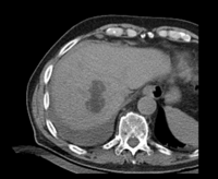
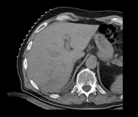
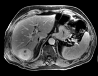
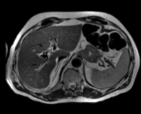
| fixed image/target: CT post | moving image 1: CT intra | moving image 2: MRI T1 pre | moving image 3: MRI T2 pre |
Objective / Background
We seek to align/fuse pre-operative MRI with intra- and post-operative CT.
Keywords
MRI, CT, IGT, intra-operative, liver, cryoablation, change detection, non-rigid registration
Input Data
- CT post reference/fixed : 0.57 x 0.57 x 5 , 512 x 512 x 37 voxels
- CT pre: 0.57 x 0.57 x 5 mm, 512 x 512 x 33 voxels
- MRI T1 pre: 1.56 x 1.56 x 2.5 mm, 256 x 256 x 88
- MRI T2 pre: 1.56 x 1.56 x 6 mm, 256 x 256 x 36
Download
Procedure
- Phase I: register CTpre - CTpost
- this registration is fairly straightforward, since FOV, contrast and initial orientation of the two scans are very similar. We perform a linear + nonrigid registration without any masking
- open the General Registration (BRAINS) module
- Fixed Image Volume: CTpost
- Moving Image Volume: CTpre
- Output Settings:
- Slicer BSpline Transform": create new transform, rename to "Xf1_Ctpre-CTpost_BSpline"
- Slicer Linear Transform: none
- Output Image Volume: create new volume, rename to "CTpre_Xf1"
- Registration Phases: check boxes for Rigid , Rigid+Scale , Affine and "BSpline"
- Main Parameters:
- B-Spline Grid Size: 7,7,3
- Leave all other settings at default
- click: Apply; runtime ~ 1 min (MacPro QuadCore 2.4GHz)
- Phase II: Create Masks
- for the MR-CT registration we have a big difference in the FOV as well as contrast and amount of anisotropy. We need to mask the liver as region of interest to stabilize the registration; otherwise the clipped FOV will cause the moving image to distort. To skip this step you may load the masks provided with the example data.
- open the Editor module
- select "CTpost" as the master volume , a new CTpost_label volume will be created.
- select the Brush tool and manually cover the liver and adjacent areas in all axial slices. This need not be accurate, the goal is to exclude the contralateral side where the FOV is clipped.
- repeat and create a similar mask for the MRI-T2.
- save results thus far
- Phase III: Affine Registration MR-CT
- this phase runs a linear registration first to better track progress. We then use this registration as input for the nonrigid registration in phase IV below. You may combine the two and include a BSpline registration phaser right away.
- open the General Registration (BRAINS) module
- Fixed Image Volume: CTpost
- Moving Image Volume: MRI_T2
- Output Settings:
- Slicer BSpline Transform": none
- Slicer Linear Transform: create new transform, rename to "Xf2_T2-CTpost_Affine"
- Output Image Volume: create new volume, rename to "MRI-T2_Xf2" (we use this for validation only)
- Registration Phases: check boxes for Rigid, "Rigid+Scale", "Affine"
- Main Parameters:
- Mask Option: select ROI button
- ROI Masking input fixed: select "CTpost_label" created in phase II above
- ROI Masking input moving: select "MRI-T2-label.nrrd" created in phase II above
- Leave all other settings at default
- click: Apply
- you should get a registration similar to the one shown in the results section below.
- Phase IV: Nonrigid Registration MR-CT
- open the General Registration (BRAINS) module
- Fixed Image Volume: CTpost
- Moving Image Volume: MRI_T2
- Output Settings:
- Slicer BSpline Transform": create new transform, rename to "Xf3_T2-CTpost_BSpline"
- Slicer Linear Transform: none
- Output Image Volume: create new volume, rename to "MRI-T2_Xf3"
- Registration Phases: check boxes for BSpline only
- Main Parameters:
- Number Of Samples: 200,000
- B-Spline Grid Size: 5,5,5
- Mask Option: select ROI button
- ROI Masking input fixed: select " "T1_mask_ROIauto" or "T1_mask_edit" edited in phase III above
- ROI Masking input moving: select "DWI_mask" created in phase I above
- Leave all other settings at default
- click: Apply
Discussion: Registration Challenges
- large differences in FOV
- strong differences in image contrast between MRI & CT
- contrast enhancement and pathology and treatment changes cause additional differences in image content
- we have strongly anisotropic voxel sizes with much less through-plane resolution