Difference between revisions of "2017 Winter Project Week/MeningiomaSegmentation"
From NAMIC Wiki
(Added examples) |
(fixed formatting) |
||
| Line 44: | Line 44: | ||
==Examples== | ==Examples== | ||
{| class="wikitable" | {| class="wikitable" | ||
| − | |Sometimes, ANTs failed to remove parts of the skull close to the tumor or wrongly removed part of the brain. | + | | |
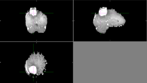
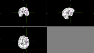
| − | [[File: | + | [[File:Case_001_ants_brain_failure.png|thumbnail|Sometimes, ANTs failed to remove parts of the skull close to the tumor or wrongly removed part of the brain.]] |
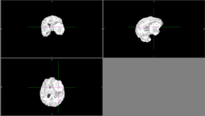
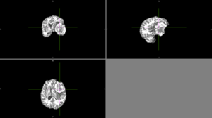
| − | + | [[File:Case_052_ants_brain.png|thumbnail|Other times, ANTs extracted the brain well.]] | |
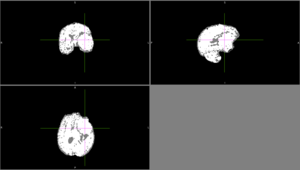
| − | [[File: | + | [[File:Case_052_ants_brain_seg.png|thumbnail|Automatic segmentation with ANTs usually classified as CSF or it classified different parts of the tumor as different types of tissue.]] |
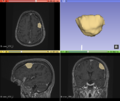
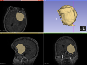
| − | Automatic segmentation with ANTs usually classified as CSF or it classified different parts of the tumor as different types of tissue. | + | [[File:Case_052_2_slicer_seg.png|thumbnail|Semi-automatic segmentation with Slicer was relatively successful. Sometimes the segmentation would bleed outside of the tumor into voxels with similar intensities.]] |
| − | [[File: | ||
| − | Semi-automatic segmentation with Slicer was relatively successful. Sometimes the segmentation would bleed outside of the tumor into voxels with similar intensities. | ||
| − | |||
| | | | ||
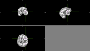
We tried FSL's FAST with different numbers of classes. None of these methods could identify the entire tumor mass as one type of tissue in this scan. | We tried FSL's FAST with different numbers of classes. None of these methods could identify the entire tumor mass as one type of tissue in this scan. | ||
| Line 58: | Line 55: | ||
[[File:Case_052_fast_5classes.png|thumbnail|FAST brain segmentation with 5 classes]] | [[File:Case_052_fast_5classes.png|thumbnail|FAST brain segmentation with 5 classes]] | ||
|} | |} | ||
| − | |||
| − | |||
Revision as of 21:09, 13 January 2017
Home < 2017 Winter Project Week < MeningiomaSegmentationKey Investigators
- Jakub Kaczmarzyk, MIT
- Satrajit Ghosh, MIT
- Omar Arnaout, Brigham and Women's Hospital
Project Description
| Objective | Approach and Plan | Progress and Next Steps |
|---|---|---|
|
|
Progress
Next steps
|
Examples
|
|
We tried FSL's FAST with different numbers of classes. None of these methods could identify the entire tumor mass as one type of tissue in this scan. |
Background and References
MR images of meningiomas that will be used in this project are available at OpenNeu.ro.

