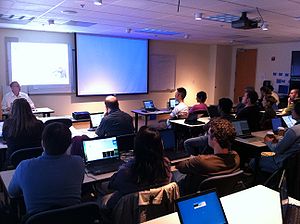
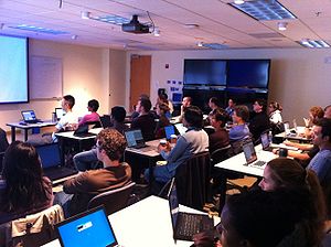
Events:UCSF-Slicer-Training-11-2010
Contents
Logistics
Location: UCSF (3rd floor) China basin campus
Date: Tuesday November 9, 2010
Registration: To sign-up for this event, please fill in the registration form and send it by e-mail to Jason Crane, PhD [jason.crane at ucsf.edu] before Friday November 5.
Tentative Agenda
- 8:45-9:00 am Computer setup assistance by the instructors
- 9:00-9:15 am Welcome and Goals of the Workshop (Ron Kikinis/Sonia Pujol)
- 9:15-9:45 am Introduction to NA-MIC and the Slicer community (Ron Kikinis)
- 9:45-10:45 am Data Loading and 3D Visualization (part1)(Sonia Pujol)
- 10:45-11:00 am Coffee break
- 11:00-11:45 am Data Loading and 3D Visualization (part 2) (Sonia Pujol)
- 11:45 am-1:00 pm Lunch
- 1:00-2:00 pm Image Registration (Sonia Pujol)
- 2:00-3:00 pm Diffusion Tensor Imaging Analysis: from DWI to DTI data (Sonia Pujol)
- 3:00-3:15 pm Coffee Break
- 3:15-4:15 pm Diffusion Tensor Imaging Analysis: from DTI data to 3D fiber bundles (Sonia Pujol)
- 4:15-4:30 pm MR Spectroscopy module in Slicer (Jason Crane)
- 4:30-5:00 pm Questions from the audience and concluding remarks
Notes on the Nrrd file format
The Nrrd file format, which is part of the NA-MIC kit, accurately represents N-dimensional raster information for scientific visualization and medical image processing. This format is used in Slicer to represent the necessary information about a DWI image volume, its anatomical orientation, and all the DWI-specific acquisition parameters for estimating the diffusion tensors, such as the measurement frame. A detailed description of the Nrrd file format use for DWI images and DTI data can be found here.
The DicomToNrrd module in Slicer can be used to convert Dicom data into Nrrd. To convert data from other file formats, such as Nifti, a tutorial describes how to construct a nhdr file from a set of images and a list of known gradients using the Teem library.
Local Organizers
- Jason Crane, PhD, Department of Radiology and Biomedical Imaging, UCSF
- Sarah Nelson, PhD, Department of Radiology and Biomedical Imaging, UCSF
- Daniel Rubin, MD, MS, Department of Radiology, Stanford University
Teaching Faculty
- Ron Kikinis, M.D., Surgical Planning Laboratory, Brigham and Women's Hospital, Harvard Medical School
- Sonia Pujol, Ph.D., Surgical Planning Laboratory, Brigham and Women's Hospital, Harvard Medical School
- Nobuhiko Hata, Ph.D., Surgical Planning Laboratory, Brigham and Women's Hospital, Harvard Medical School
Preparation for the Workshop
The workshop combines oral presentations and instructor-led hands-on sessions with the participants working on their own laptop computers. All participants are required to come with their own laptop computer and install the software and datasets prior to the event.. A minimum of 1 GB of RAM (2 GB if possible) and a dedicated graphic accelerator with 64mb of on board graphic memory are required.
Please install the Slicer3.6.1 version appropriate to the laptop computer you'll be bringing to the tutorial:
- Windows: Slicer3-3.6.1-2010-08-23-win32.exe
- Linux 64: Slicer3-3.6.1-2010-08-23-linux-x86_64
- Linux 32: Slicer3-3.6.1-2010-08-20-linux-x86
- Mac Darwin: Slicer3-3.6.1-2010-08-20-darwin-x86
Please download the 3D Visualization dataset,Registration dataset and Diffusion dataset in preparation for the workshop.
Slicer3 Training Survey
Click here to take the Slicer3 Training Survey
Back to Events 2010


